Copyright
©The Author(s) 2020.
World J Clin Cases. Aug 26, 2020; 8(16): 3411-3430
Published online Aug 26, 2020. doi: 10.12998/wjcc.v8.i16.3411
Published online Aug 26, 2020. doi: 10.12998/wjcc.v8.i16.3411
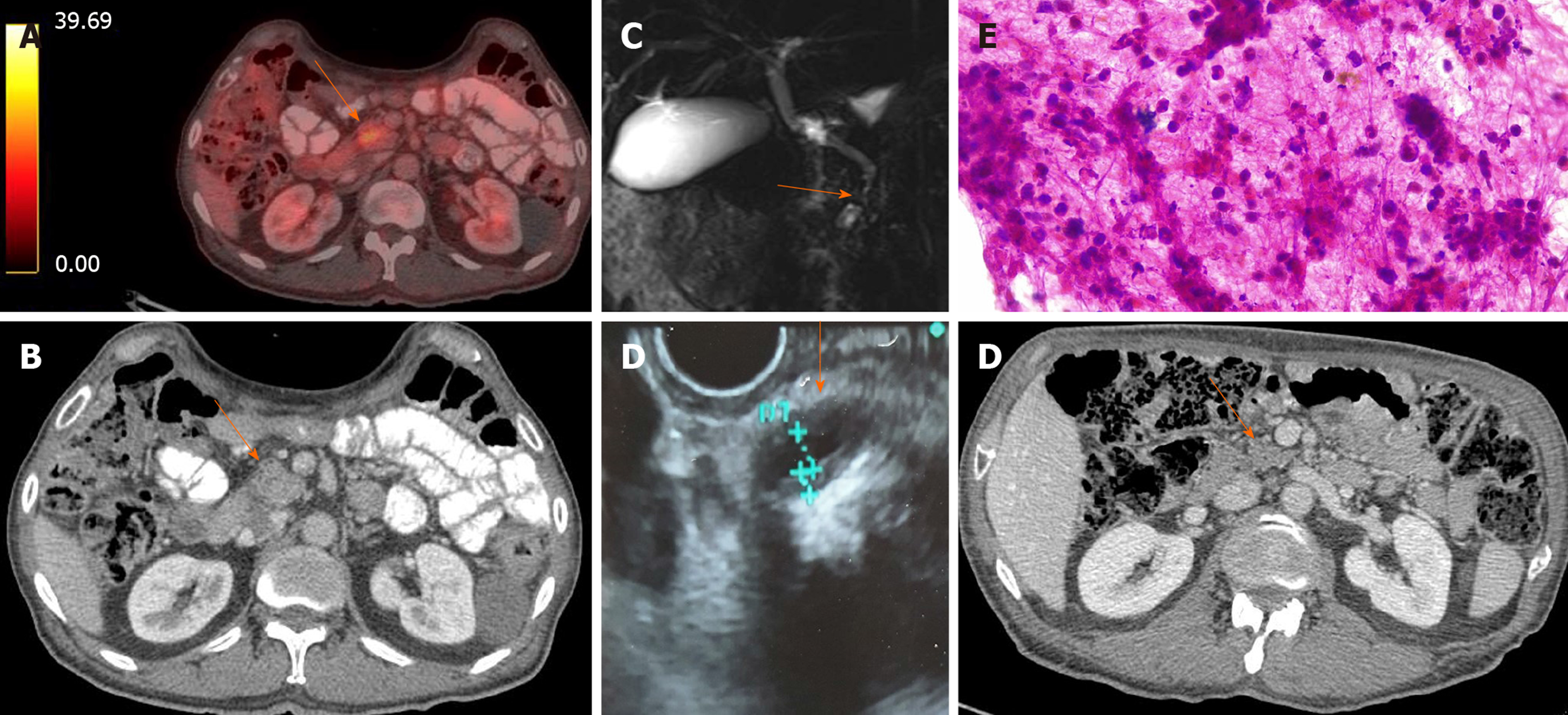
Figure 2 Idiopathic duct centric pancreatitis in a 70-year-old male with presenting with new onset diabetes mellitus patient and weight loss.
A and B: Positron emission tomography-computed tomography and simple computed tomography scan showing a heterogenous mass (arrows) in the head of the pancreas (max SUV 4.2); C: Magnetic resonance cholangiopancreatography showing a distal common bile duct common biliary duct stenosis with no proximal biliary or main pancreatic duct dilation; D: Endoscopic ultrasound showing a narrowed intrapancreatic common bile duct with symmetric hypoechoic thick walls; E: Micrography of cytology specimen from a pancreatic head mass obtained with endoscopic ultrasonography guided fine-needle aspiration, it shows a dense acute and chronic inflammatory infiltrate with numerous neutrophils in the background; F: Chest computed tomography (venous phase) showing atrophy and disappearance of the pancreatic mass in the head of the pancreas following 4 wk of steroid therapy (arrow).
- Citation: Pelaez-Luna M, Soriano-Rios A, Lira-Treviño AC, Uscanga-Domínguez L. Steroid-responsive pancreatitides. World J Clin Cases 2020; 8(16): 3411-3430
- URL: https://www.wjgnet.com/2307-8960/full/v8/i16/3411.htm
- DOI: https://dx.doi.org/10.12998/wjcc.v8.i16.3411


