Copyright
©The Author(s) 2020.
World J Clin Cases. Aug 6, 2020; 8(15): 3329-3333
Published online Aug 6, 2020. doi: 10.12998/wjcc.v8.i15.3329
Published online Aug 6, 2020. doi: 10.12998/wjcc.v8.i15.3329
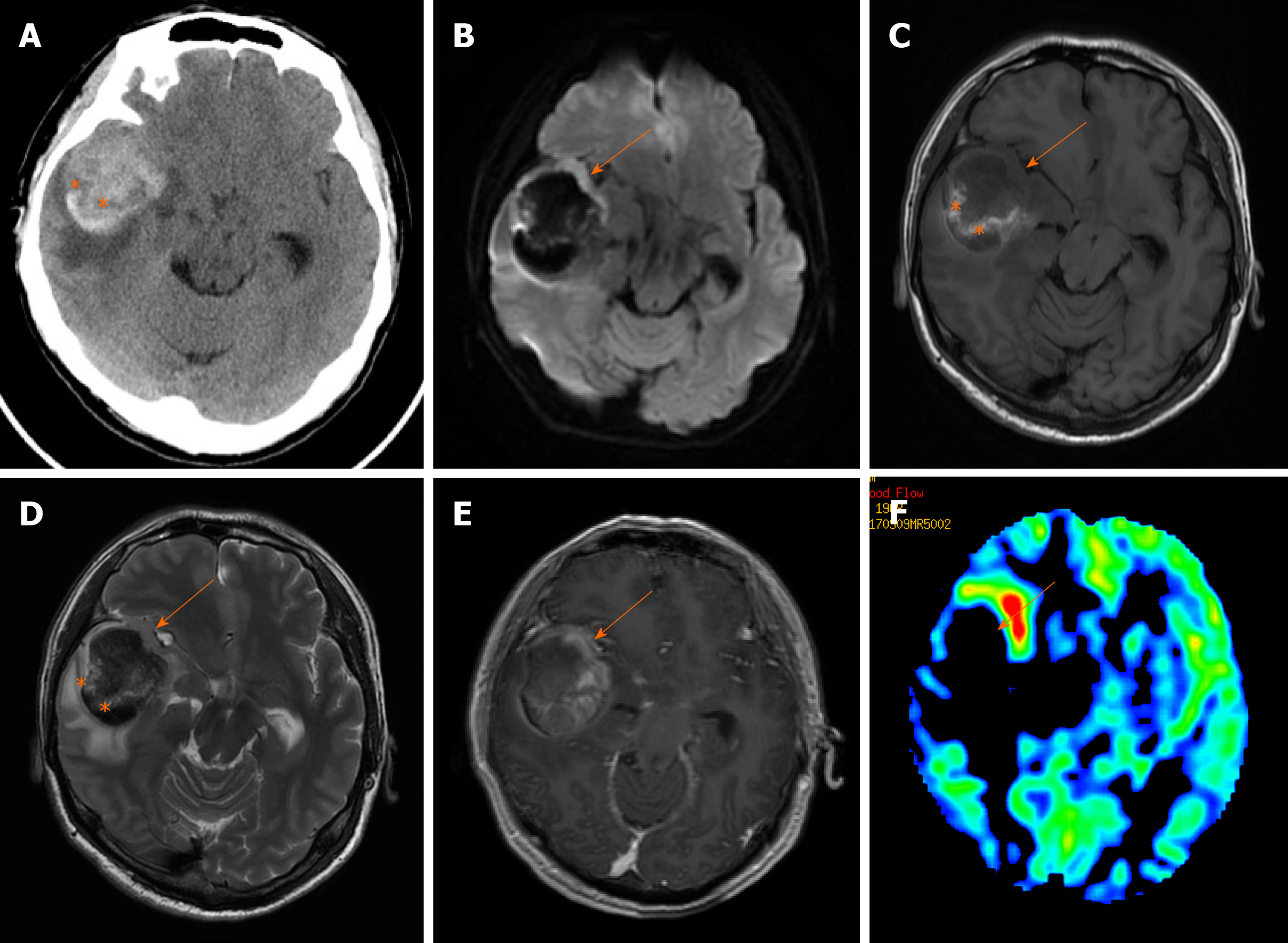
Figure 1 Brain computed tomography and magnetic resonance imaging showed a heterogeneous hyperintense signal in the right temporal lobe.
A: Non-contrast-enhanced computed tomography images showed massive hemorrhage (orange star) with perilesional edema; B: The diffusion-weighted image (b = 1000 mm/s) demonstrates a relatively hyperintense signal of the parenchyma (orange arrow); C: T1-weighted image; D: T2W image showing the parenchyma (orange arrow) of the lesion as iso- to hypointense; E: Contrast-enhanced T1-weighted image revealed a ring-like enhancing pattern; and F: Arterial spin labeling showed relatively low perfusion of the whole lesion.
- Citation: Wu YW, Zheng J, Liu LL, Cai JH, Yuan H, Ye J. Imaging of hemorrhagic primary central nervous system lymphoma: A case report. World J Clin Cases 2020; 8(15): 3329-3333
- URL: https://www.wjgnet.com/2307-8960/full/v8/i15/3329.htm
- DOI: https://dx.doi.org/10.12998/wjcc.v8.i15.3329


