Copyright
©The Author(s) 2020.
World J Clin Cases. Aug 6, 2020; 8(15): 3320-3328
Published online Aug 6, 2020. doi: 10.12998/wjcc.v8.i15.3320
Published online Aug 6, 2020. doi: 10.12998/wjcc.v8.i15.3320
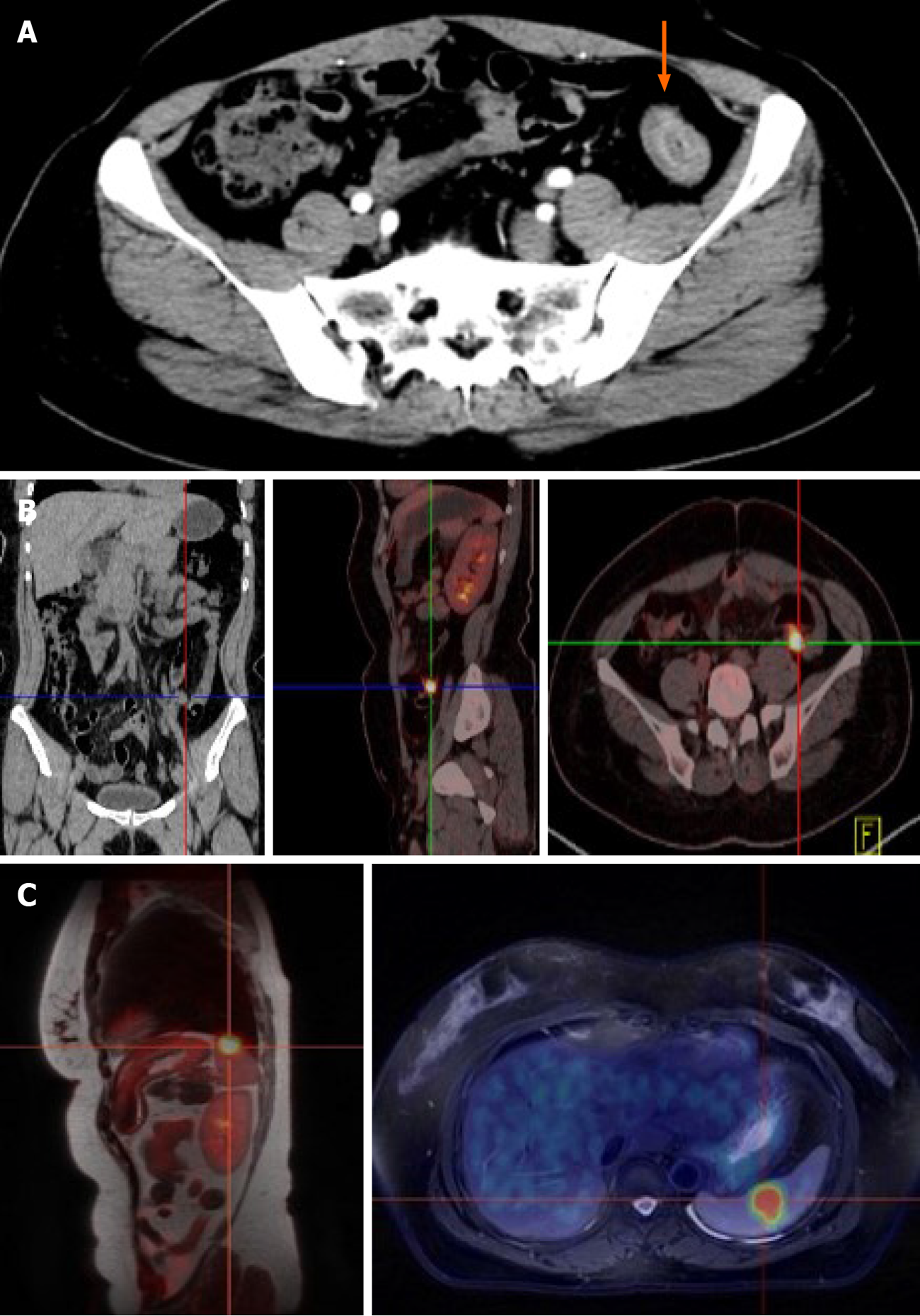
Figure 1 Imaging examinations of the colon.
A: Initial contrast-enhanced computed tomography (CT) of abdomen showed slight enhancement of the wall of sigmoid colon and a mass (arrow); B: Positron emission tomography (PET)-CT showed a high metabolic shadow in the descending colon, suggesting metastasis, but no such shadow was seen in the spleen; C: PET-magnetic resonance imaging revealed a high metabolic shadow, suggesting metastasis.
- Citation: Hu L, Zhu JY, Fang L, Yu XC, Yan ZL. Isolated metachronous splenic multiple metastases after colon cancer surgery: A case report and literature review. World J Clin Cases 2020; 8(15): 3320-3328
- URL: https://www.wjgnet.com/2307-8960/full/v8/i15/3320.htm
- DOI: https://dx.doi.org/10.12998/wjcc.v8.i15.3320


