Copyright
©The Author(s) 2020.
World J Clin Cases. Aug 6, 2020; 8(15): 3291-3298
Published online Aug 6, 2020. doi: 10.12998/wjcc.v8.i15.3291
Published online Aug 6, 2020. doi: 10.12998/wjcc.v8.i15.3291
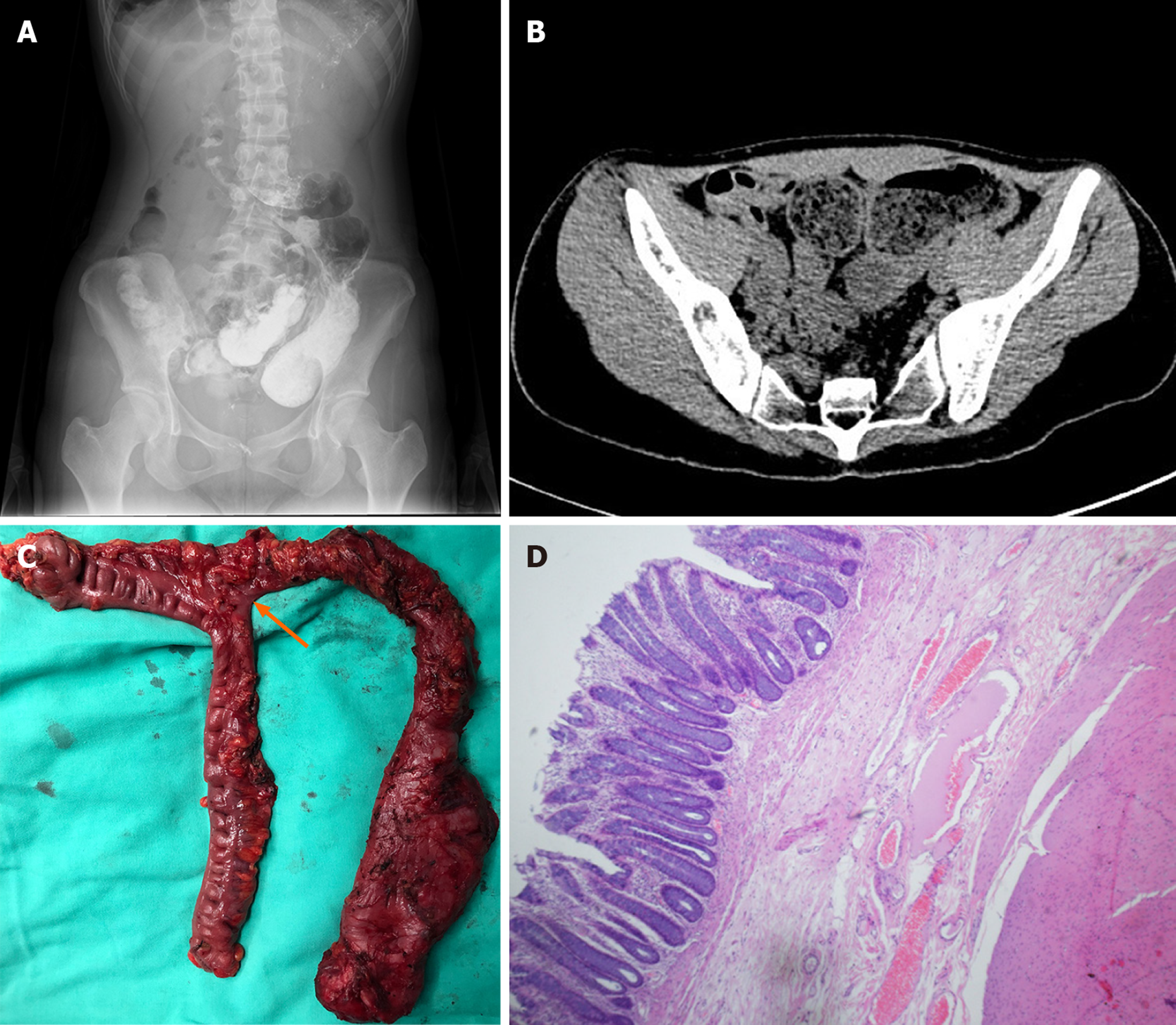
Figure 1 Related figures demonstrating the clinical characteristics of tubular colonic duplication.
A: Abdominal x-ray showed two large dilated loops filled with barium in the left lower abdomen; B: Abdominal computed tomography scan revealed two enlarged lumen with massive stored feces in the left abdominal region; C: Surgical specimen of the duplicated colon, an intestinal loop (as shown by the arrow) was separated from the transverse colon adjacent to the splenic flexure and extended to the left iliac fossa with a dead end; D: Histopathologic evaluation revealed normal alimentary structures with well-formed mucosa and smooth muscular layer, which further confirmed the diagnosis.
- Citation: Li GB, Han JG, Wang ZJ, Zhai ZW, Tao Y. Successful management of tubular colonic duplication using a laparoscopic approach: A case report and review of the literature. World J Clin Cases 2020; 8(15): 3291-3298
- URL: https://www.wjgnet.com/2307-8960/full/v8/i15/3291.htm
- DOI: https://dx.doi.org/10.12998/wjcc.v8.i15.3291


