Copyright
©The Author(s) 2020.
World J Clin Cases. Aug 6, 2020; 8(15): 3267-3279
Published online Aug 6, 2020. doi: 10.12998/wjcc.v8.i15.3267
Published online Aug 6, 2020. doi: 10.12998/wjcc.v8.i15.3267
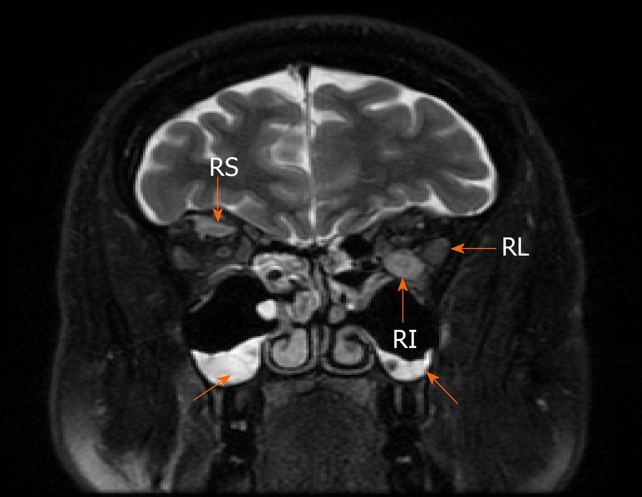
Figure 2 Magnetic resonance imaging of the orbital cavity.
1.5-T Magnetic resonance imaging of the orbital cavity showing the swelling of the extra orbital muscles and sinusitis (T2-weighed image) (arrows): RL – musculus rectus lateralis sinister [5.5 mm (normal range 3.3 mm)], RI – musculus rectus inferior sinister [8 mm (normal range 4.6 mm)], RS – musculus rectus superior dexter [5 mm (normal range 4.6 mm)].
- Citation: Strainiene S, Sarlauskas L, Savlan I, Liakina V, Stundiene I, Valantinas J. Multi-organ IgG4-related disease continues to mislead clinicians: A case report and literature review. World J Clin Cases 2020; 8(15): 3267-3279
- URL: https://www.wjgnet.com/2307-8960/full/v8/i15/3267.htm
- DOI: https://dx.doi.org/10.12998/wjcc.v8.i15.3267


