Copyright
©The Author(s) 2020.
World J Clin Cases. Aug 6, 2020; 8(15): 3177-3187
Published online Aug 6, 2020. doi: 10.12998/wjcc.v8.i15.3177
Published online Aug 6, 2020. doi: 10.12998/wjcc.v8.i15.3177
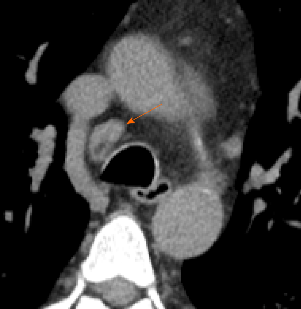
Figure 9 Computed tomography images of lymphadenopathy.
61-year man admitted to the emergency room presenting cough and dyspnea. Chest computed tomography showed a lymphadenopathy (short axis > 10 mm) in right lower paratracheal station (orange arrow).
- Citation: Caruso D, Polidori T, Guido G, Nicolai M, Bracci B, Cremona A, Zerunian M, Polici M, Pucciarelli F, Rucci C, Dominicis CD, Girolamo MD, Argento G, Sergi D, Laghi A. Typical and atypical COVID-19 computed tomography findings. World J Clin Cases 2020; 8(15): 3177-3187
- URL: https://www.wjgnet.com/2307-8960/full/v8/i15/3177.htm
- DOI: https://dx.doi.org/10.12998/wjcc.v8.i15.3177


