Copyright
©The Author(s) 2020.
World J Clin Cases. Aug 6, 2020; 8(15): 3164-3176
Published online Aug 6, 2020. doi: 10.12998/wjcc.v8.i15.3164
Published online Aug 6, 2020. doi: 10.12998/wjcc.v8.i15.3164
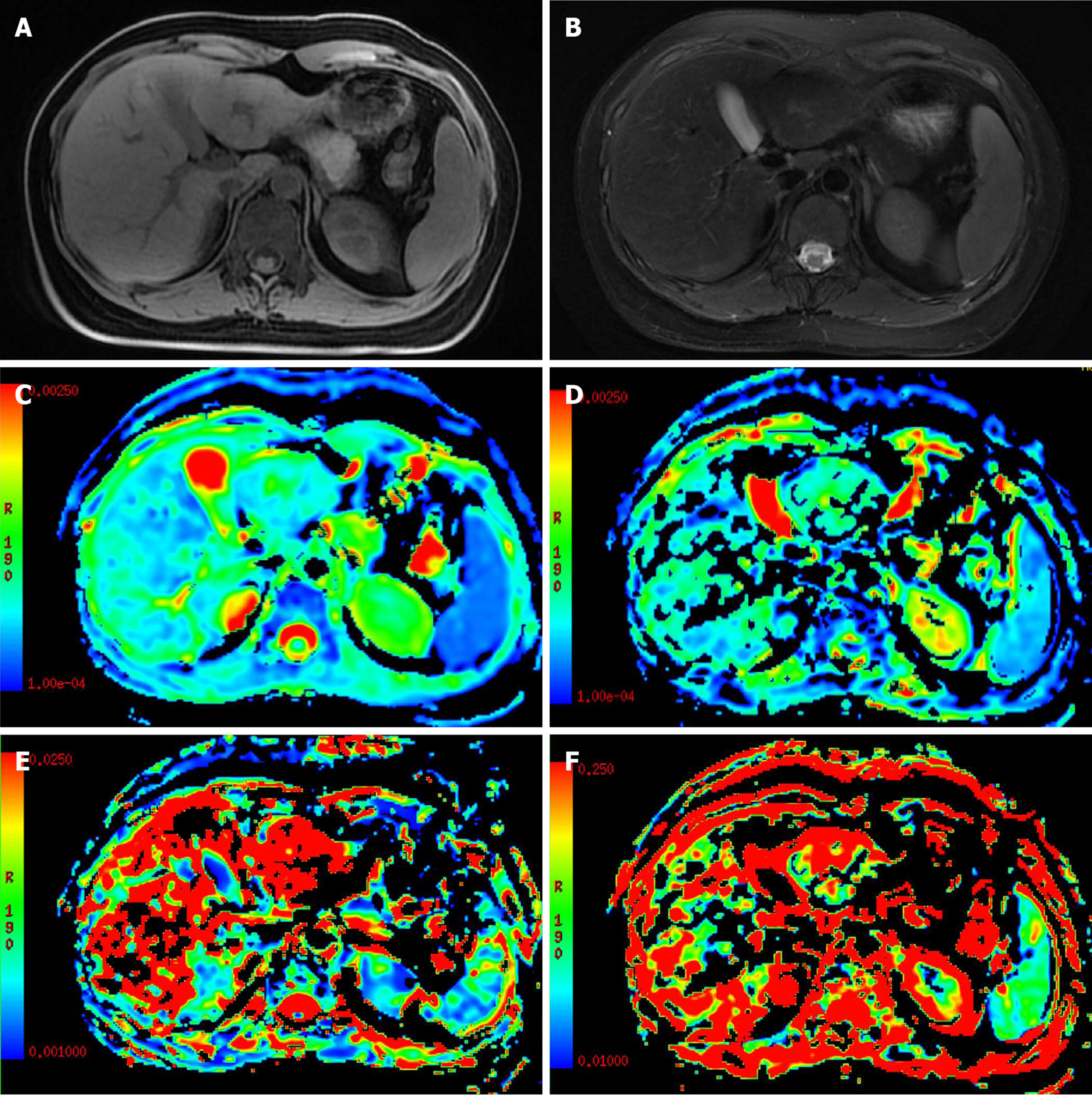
Figure 2 A 28-year-old female patient with focal nodular hyperplasia in the left lobe of the liver.
The tumor was isointense on T1-weighted image (T1WI) and T2-weighted image (T2WI) with a star-shaped area in the center of the lesion (hypointense on T1WI and hyperintense on T2WI). A: T1WI; B: T2WI; C: ADC map; D: D map; E: D* map; F: F map.
- Citation: Tao YY, Zhou Y, Wang R, Gong XQ, Zheng J, Yang C, Yang L, Zhang XM. Progress of intravoxel incoherent motion diffusion-weighted imaging in liver diseases. World J Clin Cases 2020; 8(15): 3164-3176
- URL: https://www.wjgnet.com/2307-8960/full/v8/i15/3164.htm
- DOI: https://dx.doi.org/10.12998/wjcc.v8.i15.3164


