Copyright
©The Author(s) 2020.
World J Clin Cases. Jul 26, 2020; 8(14): 3122-3129
Published online Jul 26, 2020. doi: 10.12998/wjcc.v8.i14.3122
Published online Jul 26, 2020. doi: 10.12998/wjcc.v8.i14.3122
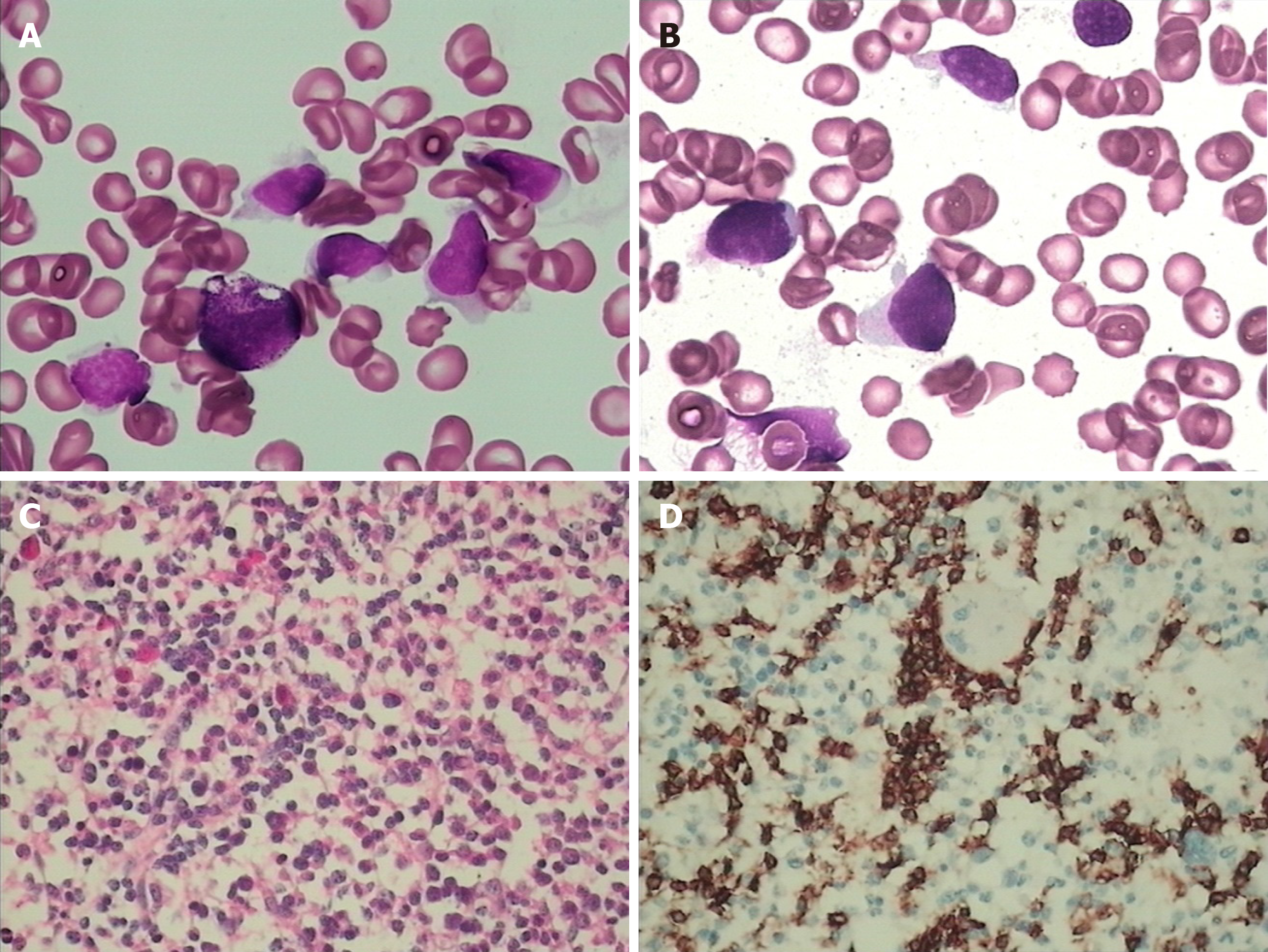
Figure 3 Morphology and immunohistochemistry analysis of bone marrow and peripheral blood.
A and B: Bone marrow aspirate smear (panel A) and blood smear (panel B) show atypical lymphocytes with round or irregular shape, less cytoplasm, irregular nuclear contours, and visible nucleoli (Wright-Giemsa, 1000 ×, oil); C and D: Bone marrow biopsy showed many atypical lymphocyte-infiltrated sinuses (panel C, hematoxylin and eosin staining, 400 ×) were positive for CD3 (panel C, 400 ×).
- Citation: Wang XT, Guo W, Sun M, Han W, Du ZH, Wang XX, Du BB, Bai O. Effect of chidamide on treating hepatosplenic T-cell lymphoma: A case report. World J Clin Cases 2020; 8(14): 3122-3129
- URL: https://www.wjgnet.com/2307-8960/full/v8/i14/3122.htm
- DOI: https://dx.doi.org/10.12998/wjcc.v8.i14.3122


