Copyright
©The Author(s) 2020.
World J Clin Cases. Jul 26, 2020; 8(14): 3114-3121
Published online Jul 26, 2020. doi: 10.12998/wjcc.v8.i14.3114
Published online Jul 26, 2020. doi: 10.12998/wjcc.v8.i14.3114
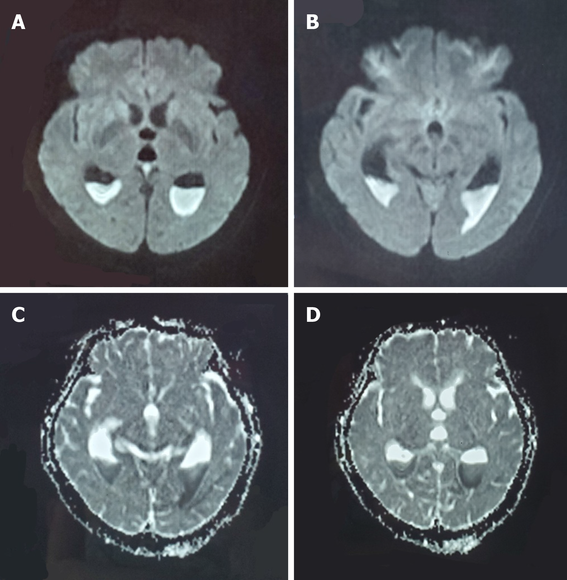
Figure 1 Head magnetic resonance imaging before admission.
A and B: The fester-traditional cerebrospinal fluid level was seen inside the posterior horns of the bilateral lateral ventricles, and the fester diffuse weighted imaging showed a high signal; C and D: The fester showed a low signal on the apparent diffusion coefficient map.
- Citation: Xue H, Wang XH, Shi L, Wei Q, Zhang YM, Yang HF. Dental focal infection-induced ventricular and spinal canal empyema: A case report. World J Clin Cases 2020; 8(14): 3114-3121
- URL: https://www.wjgnet.com/2307-8960/full/v8/i14/3114.htm
- DOI: https://dx.doi.org/10.12998/wjcc.v8.i14.3114


