Copyright
©The Author(s) 2020.
World J Clin Cases. Jul 26, 2020; 8(14): 3108-3113
Published online Jul 26, 2020. doi: 10.12998/wjcc.v8.i14.3108
Published online Jul 26, 2020. doi: 10.12998/wjcc.v8.i14.3108
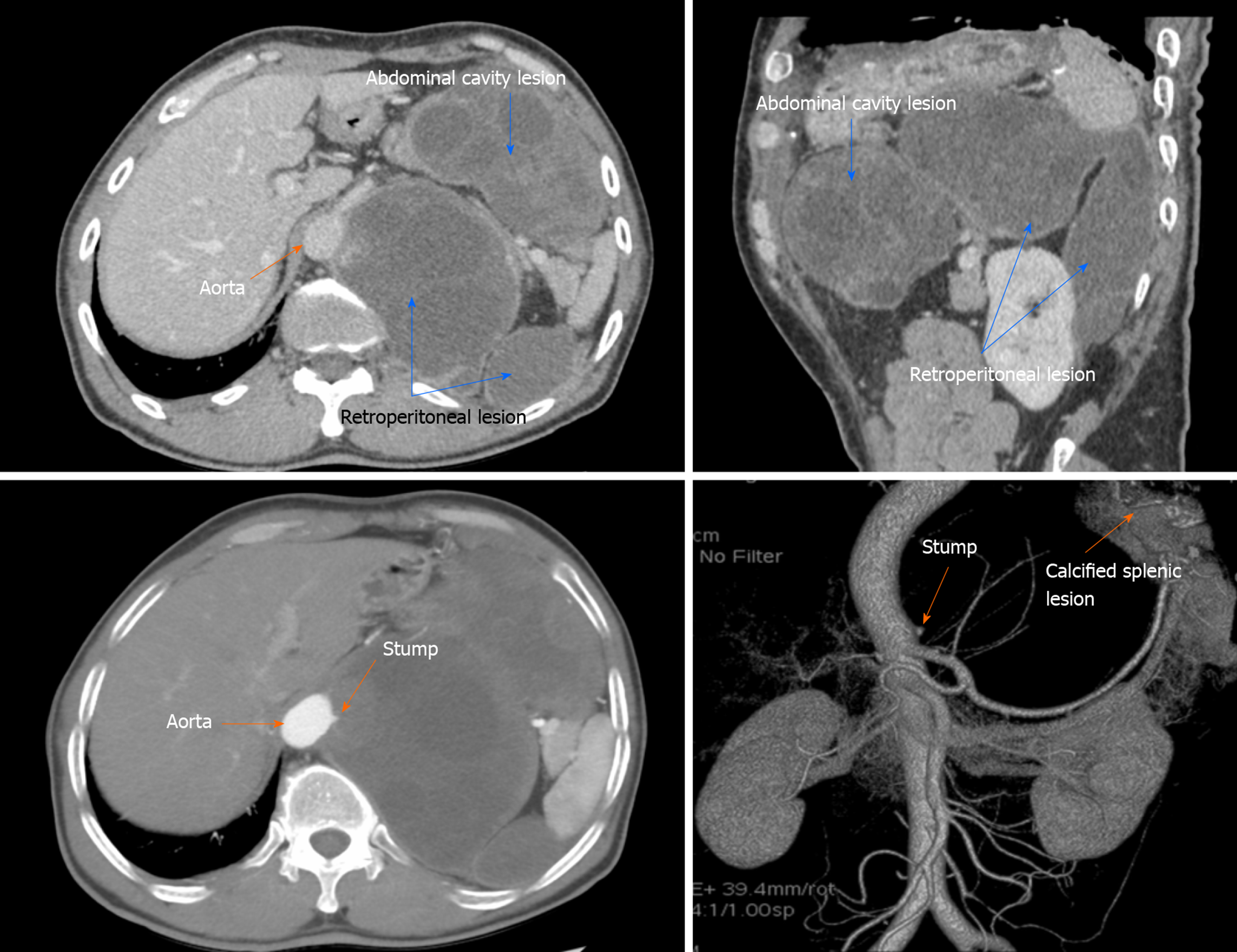
Figure 1 Preoperative computed tomography and angiography.
A and B: Computed tomography scan showed a lesion in the abdominal cavity and a retroperitoneal lesion, which was firmly attached to the abdominal aortic wall; C: At the arterial phase, an aortic branch was found to be buried in the lesion with a dead end; D: Angiography showed further evidence of the dead-end arterial branch of the abdominal aorta, and calcified splenic lesion after the initial splenic cystic echinococcosis surgery.
- Citation: Taxifulati N, Yang XA, Zhang XF, Aini A, Abulizi A, Ma X, Abulati A, Wang F, Xu K, Aji T, Shao YM, Ahan A. Multiple recurrent cystic echinococcosis with abdominal aortic involvement: A case report. World J Clin Cases 2020; 8(14): 3108-3113
- URL: https://www.wjgnet.com/2307-8960/full/v8/i14/3108.htm
- DOI: https://dx.doi.org/10.12998/wjcc.v8.i14.3108


