Copyright
©The Author(s) 2020.
World J Clin Cases. Jul 26, 2020; 8(14): 3064-3073
Published online Jul 26, 2020. doi: 10.12998/wjcc.v8.i14.3064
Published online Jul 26, 2020. doi: 10.12998/wjcc.v8.i14.3064
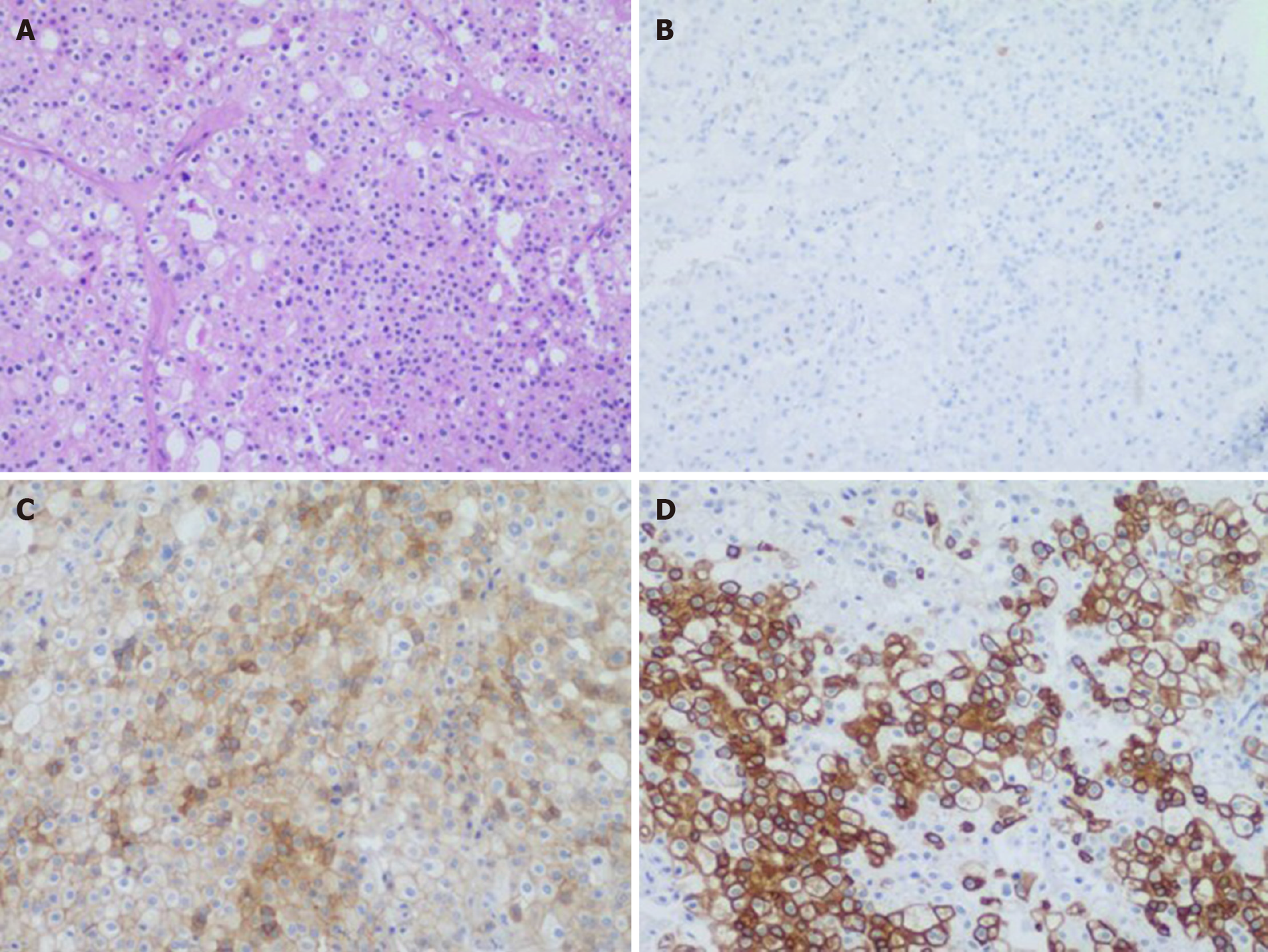
Figure 6 A 50-year-old male with pathologically proven chromophobe renal cell carcinoma in the left kidney.
A: Hematoxylin and eosin staining showing tumor cells were arranged in solid nests and gland pattern with thick-walled blood vessels, clear cell membrane, reticulated cytoplasm, and “raisinoid” nuclear membranes (100 ×); B: Immunohistochemical staining showing negative expression of carbonic anhydrase 9 (100 ×); C: Cluster of differentiation 117 was moderately diffusely positive in the cytoplasm/membrane (100 ×); D: Cytokeratin 7 was strongly diffusely positive in the cytoplasm/membrane (100 ×).
- Citation: Yang F, Zhao ZC, Hu AJ, Sun PF, Zhang B, Yu MC, Wang J. Synchronous sporadic bilateral multiple chromophobe renal cell carcinoma accompanied by a clear cell carcinoma and a cyst: A case report. World J Clin Cases 2020; 8(14): 3064-3073
- URL: https://www.wjgnet.com/2307-8960/full/v8/i14/3064.htm
- DOI: https://dx.doi.org/10.12998/wjcc.v8.i14.3064


