Copyright
©The Author(s) 2020.
World J Clin Cases. Jul 26, 2020; 8(14): 3064-3073
Published online Jul 26, 2020. doi: 10.12998/wjcc.v8.i14.3064
Published online Jul 26, 2020. doi: 10.12998/wjcc.v8.i14.3064
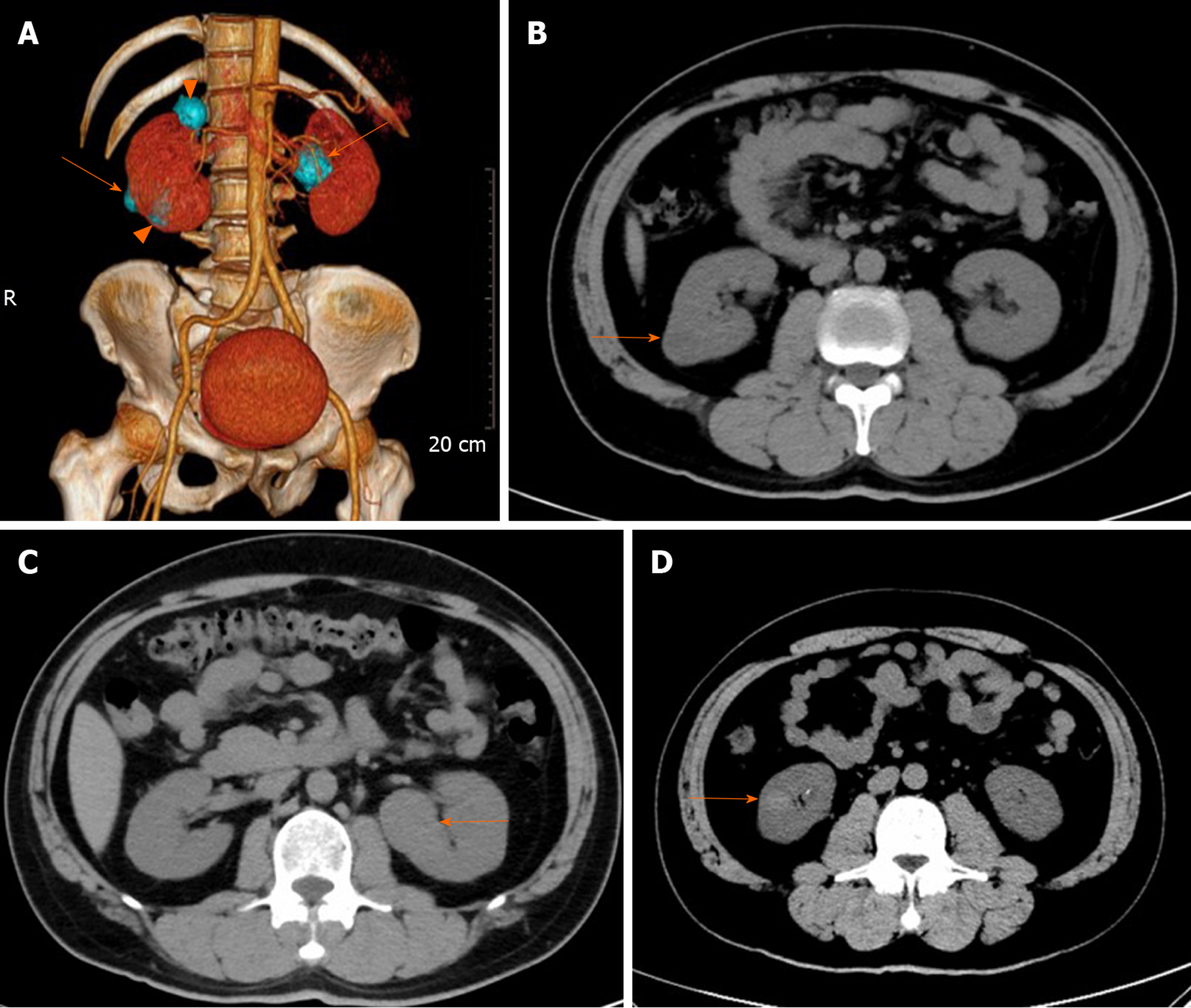
Figure 1 Computed tomography imaging of the patient.
A: Volume representation shows bilateral multiple renal tumors (4 masses); B: Pre-operative axial computed tomography (CT) imaging sections showing a 1.9 cm × 1.9 cm × 2.0 cm tumor arising from the right kidney, exophytic, heterogeneous hypodense, without calcification and hemorrhage, proven to be chromophobe renal cell carcinoma (orange arrow); C: Axial CT imaging sections showing a 3.3 cm × 3.2 cm × 2.9 cm tumor arising from the left kidney, exophytic, homogeneous isodense, without calcification and hemorrhage, proven to be a chromophobe renal cell carcinoma (orange arrow); and D: Axial CT imaging sections showing a 1.4 cm × 1.3 cm × 1.1 cm tumor arising from the right kidney, homogeneous hyperdense, without calcification and hemorrhage, proven to be a clear cell renal cell carcinoma (orange arrow).
- Citation: Yang F, Zhao ZC, Hu AJ, Sun PF, Zhang B, Yu MC, Wang J. Synchronous sporadic bilateral multiple chromophobe renal cell carcinoma accompanied by a clear cell carcinoma and a cyst: A case report. World J Clin Cases 2020; 8(14): 3064-3073
- URL: https://www.wjgnet.com/2307-8960/full/v8/i14/3064.htm
- DOI: https://dx.doi.org/10.12998/wjcc.v8.i14.3064


