Copyright
©The Author(s) 2020.
World J Clin Cases. Jul 26, 2020; 8(14): 3039-3049
Published online Jul 26, 2020. doi: 10.12998/wjcc.v8.i14.3039
Published online Jul 26, 2020. doi: 10.12998/wjcc.v8.i14.3039
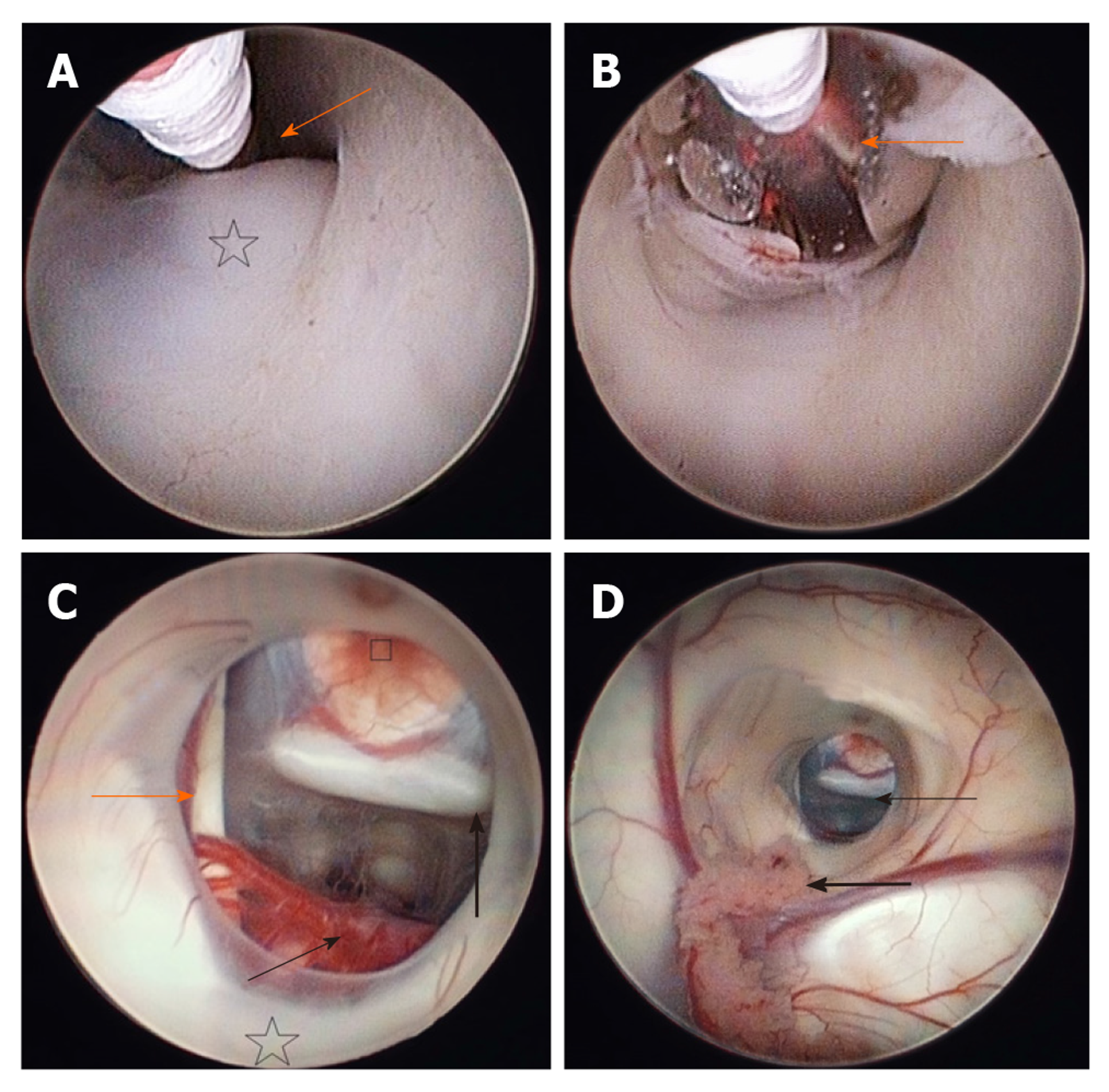
Figure 3 The floor of the third ventricle.
A: The tip of the Fogarty balloon is seen, located in the area of the thin, translucent membrane, just in front of the mammillary bodies (star), which is the target for the fenestration (arrow); B: The fenestration is underway. The floor of the third ventricle has just ben perforated and the opening is being enlarged with a Fogarty balloon. The vessels of the posterior circulation can be seen through the translucent wall of the inflated balloon (arrow); C: The ventriculostomy after the completed fenestration. The posterior circulation (the basilar artery with its bifurcation into posterior cerebral arteries and their branches) can be seen posteriorly (thin arrow), anteriorly is the clivus, the dorsum sellae above (thick arrow) and the tuber cinereum (square). The 3rd cranial nerve is running on the left (orange arrow). The furthest behind, just above the fenestration, mammillary bodies and thalami are located (star); D: The fenestration seen from the lateral ventricle through the foramen of Monro. In the opening, the clivus can be clearly seen (thin arrow) and the tuber cinereum in the front. The choroid plexus is located at the bottom of the figure (thick arrow).
- Citation: Munda M, Spazzapan P, Bosnjak R, Velnar T. Endoscopic third ventriculostomy in obstructive hydrocephalus: A case report and analysis of operative technique. World J Clin Cases 2020; 8(14): 3039-3049
- URL: https://www.wjgnet.com/2307-8960/full/v8/i14/3039.htm
- DOI: https://dx.doi.org/10.12998/wjcc.v8.i14.3039


