Copyright
©The Author(s) 2020.
World J Clin Cases. Jul 26, 2020; 8(14): 3039-3049
Published online Jul 26, 2020. doi: 10.12998/wjcc.v8.i14.3039
Published online Jul 26, 2020. doi: 10.12998/wjcc.v8.i14.3039
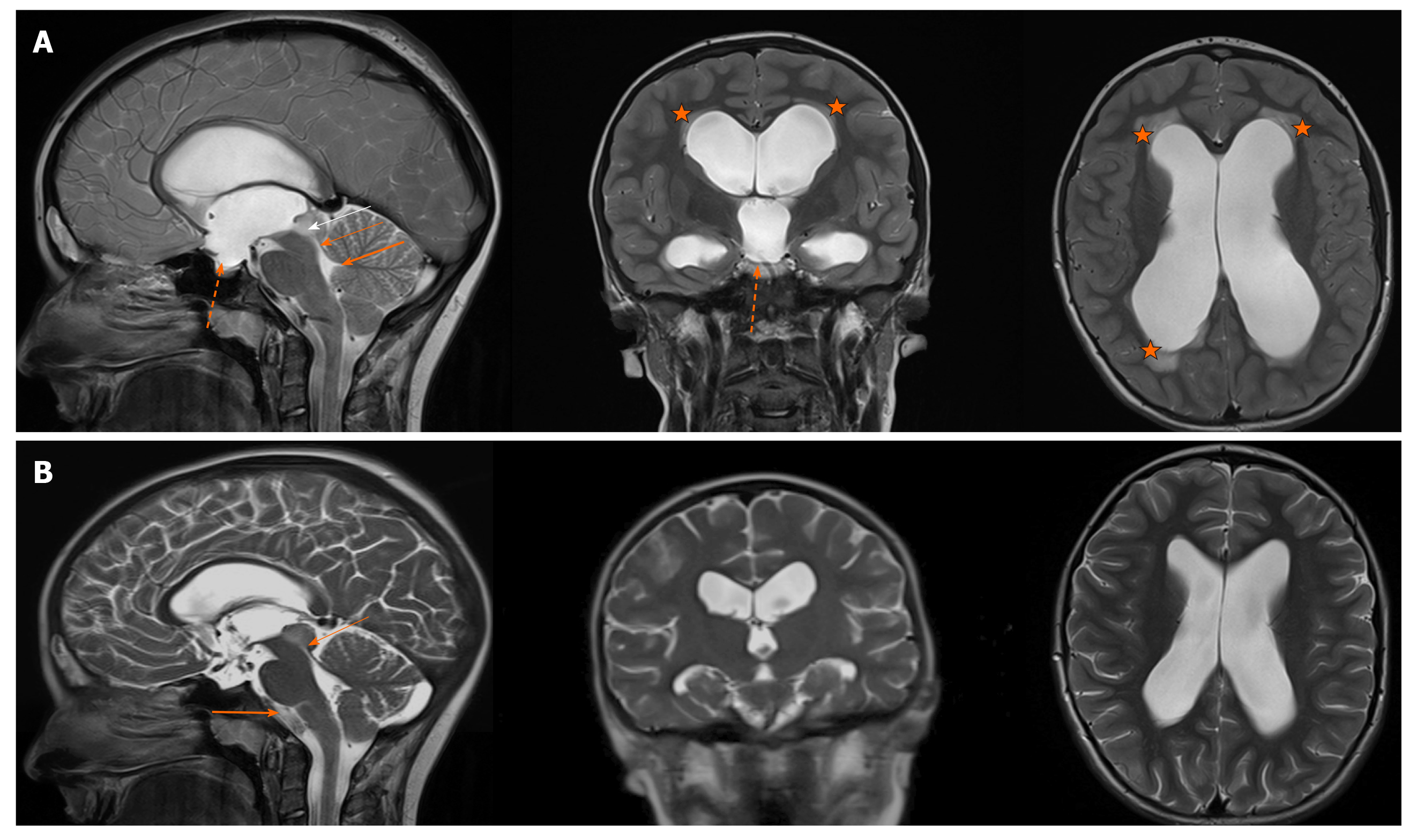
Figure 1 The T2-phases of the magnetic resonance imaging (sagittal, coronal and axial views) revealing a tumour in the right part of the tectum (white arrow), obstructing the aqueduct of Sylvius and causing extensive hydrocephalus.
A: The enlarged third ventricle is bulging into the sella turcica (dotted arrow). Periventricular lucences can also be seen (star). The thick arrow indicates the stenosis, the thin arrow indicates a normal fourth ventricle width. B: After one year, the control MRI (T2-phase) showed the regression of ventriculomegaly as a result of the endoscopic third ventriculostomy (ETV). The stenosis in the aqueduct is still present to the same degree (thin arrow). The cerebrospinal fluid flow artefact in the third ventricle can be seen (thick arrow), confirming the third ventricle floor opening after the successful ETV. The tumour has not been progressing. The supratentorial ventricles are narrower.
- Citation: Munda M, Spazzapan P, Bosnjak R, Velnar T. Endoscopic third ventriculostomy in obstructive hydrocephalus: A case report and analysis of operative technique. World J Clin Cases 2020; 8(14): 3039-3049
- URL: https://www.wjgnet.com/2307-8960/full/v8/i14/3039.htm
- DOI: https://dx.doi.org/10.12998/wjcc.v8.i14.3039


