Copyright
©The Author(s) 2020.
World J Clin Cases. Jul 26, 2020; 8(14): 3031-3038
Published online Jul 26, 2020. doi: 10.12998/wjcc.v8.i14.3031
Published online Jul 26, 2020. doi: 10.12998/wjcc.v8.i14.3031
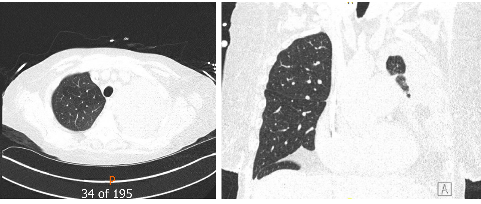
Figure 3 Computed tomography chest (Axial and coronal reformatted images) show a large left pleural effusion with near-complete opacification of the left lung parenchyma.
- Citation: Deshwal H, Ghosh S, Hogan K, Akindipe O, Lane CR, Mehta AC. Spontaneous pneumothorax in a single lung transplant recipient-a blessing in disguise: A case report. World J Clin Cases 2020; 8(14): 3031-3038
- URL: https://www.wjgnet.com/2307-8960/full/v8/i14/3031.htm
- DOI: https://dx.doi.org/10.12998/wjcc.v8.i14.3031


