Copyright
©The Author(s) 2020.
World J Clin Cases. Jul 26, 2020; 8(14): 2902-2916
Published online Jul 26, 2020. doi: 10.12998/wjcc.v8.i14.2902
Published online Jul 26, 2020. doi: 10.12998/wjcc.v8.i14.2902
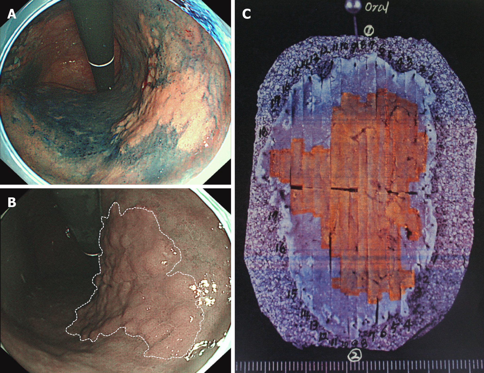
Figure 6 Narrow-band imaging endoscopy for determining the horizontal margin of gastric dysplasia before endoscopic submucosal dissection.
A: Conventional chromoendoscopy using indigo carmine is useful for determining the horizontal margin of gastric neoplasia. However, this procedure requires a dye solution and is time-consuming; B: When the endoscopist presses the button, narrow-band imaging (NBI) endoscopy can be easily performed as a virtual chromoendoscopy. The tumor margin is evident between the large brownish lesion and greenish background mucosa (dotted white line); C: An en bloc-resected specimen by endoscopic submucosal dissection. The orange-colored area indicates a tubulovillous adenoma of 46 mm × 36 mm size, corresponding to the tumor extent determined by NBI endoscopy.
- Citation: Cho JH, Jeon SR, Jin SY. Clinical applicability of gastroscopy with narrow-band imaging for the diagnosis of Helicobacter pylori gastritis, precancerous gastric lesion, and neoplasia. World J Clin Cases 2020; 8(14): 2902-2916
- URL: https://www.wjgnet.com/2307-8960/full/v8/i14/2902.htm
- DOI: https://dx.doi.org/10.12998/wjcc.v8.i14.2902


