Copyright
©The Author(s) 2020.
World J Clin Cases. Jul 26, 2020; 8(14): 2902-2916
Published online Jul 26, 2020. doi: 10.12998/wjcc.v8.i14.2902
Published online Jul 26, 2020. doi: 10.12998/wjcc.v8.i14.2902
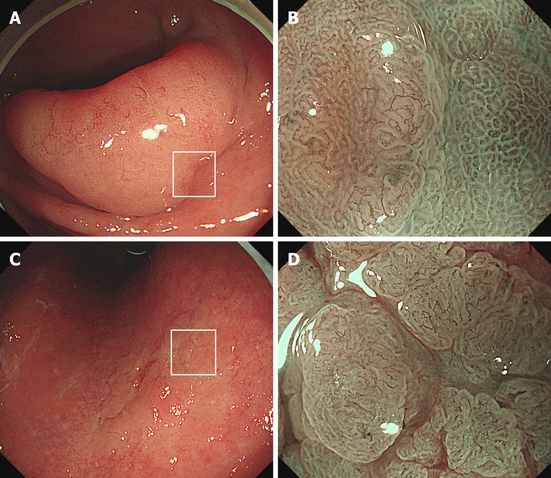
Figure 5 White-light endoscopy and magnifying narrow-band imaging images of high grade dysplasia.
A: An elevated lesion (40 mm × 30 mm) at the gastric antrum; B: Brownish area showing an irregular mucosal surface and a microvascular pattern (left side), indicating a high grade dysplasia. The demarcation line is evident between the dysplasia and background mucosa with intestinal metaplasia (also see white box in A); C: A slightly elevated lesion (35 mm × 20 mm) at the lesser curvature of the gastric corpus; D: The microvessels within irregular, nodular lesion show tortuosity and variation in shape. This magnifying narrow-band imaging endoscopic finding indicates high grade dysplasia (also see white box in C).
- Citation: Cho JH, Jeon SR, Jin SY. Clinical applicability of gastroscopy with narrow-band imaging for the diagnosis of Helicobacter pylori gastritis, precancerous gastric lesion, and neoplasia. World J Clin Cases 2020; 8(14): 2902-2916
- URL: https://www.wjgnet.com/2307-8960/full/v8/i14/2902.htm
- DOI: https://dx.doi.org/10.12998/wjcc.v8.i14.2902


