Copyright
©The Author(s) 2020.
World J Clin Cases. Jul 26, 2020; 8(14): 2902-2916
Published online Jul 26, 2020. doi: 10.12998/wjcc.v8.i14.2902
Published online Jul 26, 2020. doi: 10.12998/wjcc.v8.i14.2902
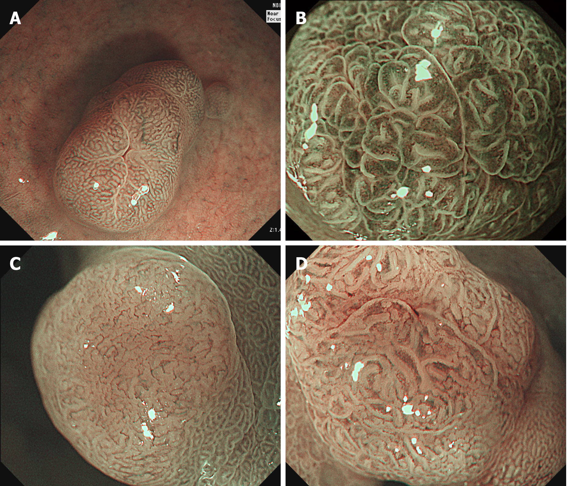
Figure 4 Magnifying narrow-band imaging findings of microvascular patterns for diagnosing the gastric polypoid lesions.
A: Honeycomb-like pattern (fundic gland polyp); B: Dense vascular pattern (hyperplastic polyp); C: Fine network within a light brown area (low grade dysplasia); D: Core vascular pattern (low grade dysplasia).
- Citation: Cho JH, Jeon SR, Jin SY. Clinical applicability of gastroscopy with narrow-band imaging for the diagnosis of Helicobacter pylori gastritis, precancerous gastric lesion, and neoplasia. World J Clin Cases 2020; 8(14): 2902-2916
- URL: https://www.wjgnet.com/2307-8960/full/v8/i14/2902.htm
- DOI: https://dx.doi.org/10.12998/wjcc.v8.i14.2902


