Copyright
©The Author(s) 2020.
World J Clin Cases. Jul 26, 2020; 8(14): 2902-2916
Published online Jul 26, 2020. doi: 10.12998/wjcc.v8.i14.2902
Published online Jul 26, 2020. doi: 10.12998/wjcc.v8.i14.2902
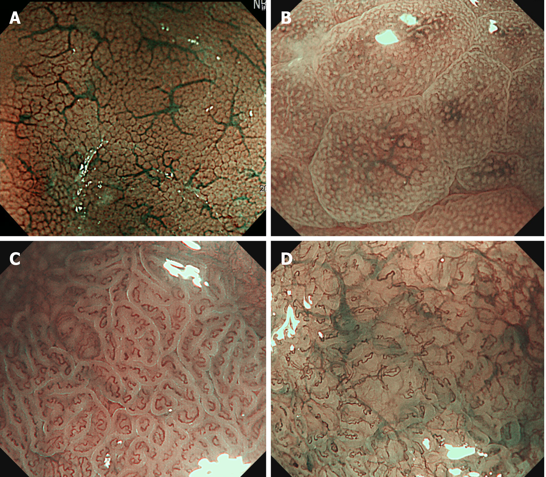
Figure 2 Magnifying narrow-band imaging endoscopy images of Helicobacter pylori-positive corpus mucosa and atrophic gastritis.
A: Normal mucosal pattern showing a honeycomb-like subepithelial capillary network and regular arrangement of collecting venules; B: Helicobacter pylori-infected status presenting as polygonal swollen mucosa with dilated round crypt opening; C: Ridged surface structures encasing dilated coiled subepithelial capillaries indicate the presence of atrophic gastritis; D: Severe mucosal atrophy characterized by irregular coiled microvessels, loss of gastric pits, and greenish submucosal vessels.
- Citation: Cho JH, Jeon SR, Jin SY. Clinical applicability of gastroscopy with narrow-band imaging for the diagnosis of Helicobacter pylori gastritis, precancerous gastric lesion, and neoplasia. World J Clin Cases 2020; 8(14): 2902-2916
- URL: https://www.wjgnet.com/2307-8960/full/v8/i14/2902.htm
- DOI: https://dx.doi.org/10.12998/wjcc.v8.i14.2902


