Copyright
©The Author(s) 2020.
World J Clin Cases. Jul 6, 2020; 8(13): 2885-2892
Published online Jul 6, 2020. doi: 10.12998/wjcc.v8.i13.2885
Published online Jul 6, 2020. doi: 10.12998/wjcc.v8.i13.2885
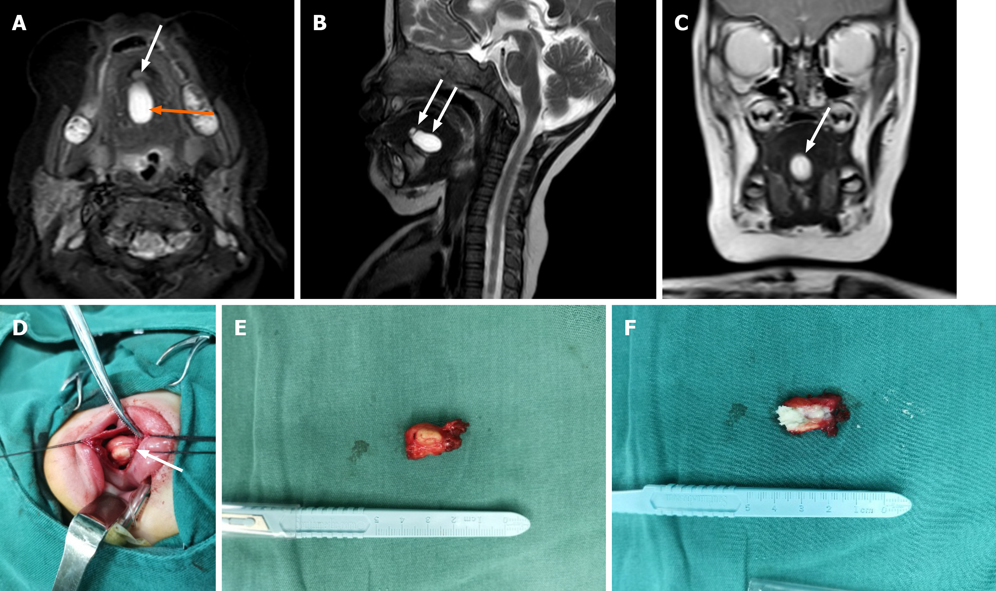
Figure 4 Infant during second surgery.
A: Second preoperative magnetic resonance imaging of large mass (orange arrow, 2.0 cm × 1.5 cm) midline floor of the mouth with high signal intensity and tiny suspect lesion (white arrow); B: Magnetic resonance imaging (sagittal plane) showing large high-signal (wide arrow) and tiny suspect (narrow arrow) lesions; C: Magnetic resonance imaging (coronal plane) of one lesion only; D: Completely encapsulated mass; E: Fully excised lesion (2.0 cm × 1.0 cm); F: Cross section with keratin content.
- Citation: Liu NN, Zhang XY, Tang YY, Wang ZM. Two sequential surgeries in infant with multiple floor of the mouth dermoid cysts: A case report. World J Clin Cases 2020; 8(13): 2885-2892
- URL: https://www.wjgnet.com/2307-8960/full/v8/i13/2885.htm
- DOI: https://dx.doi.org/10.12998/wjcc.v8.i13.2885


