Copyright
©The Author(s) 2020.
World J Clin Cases. Jul 6, 2020; 8(13): 2885-2892
Published online Jul 6, 2020. doi: 10.12998/wjcc.v8.i13.2885
Published online Jul 6, 2020. doi: 10.12998/wjcc.v8.i13.2885
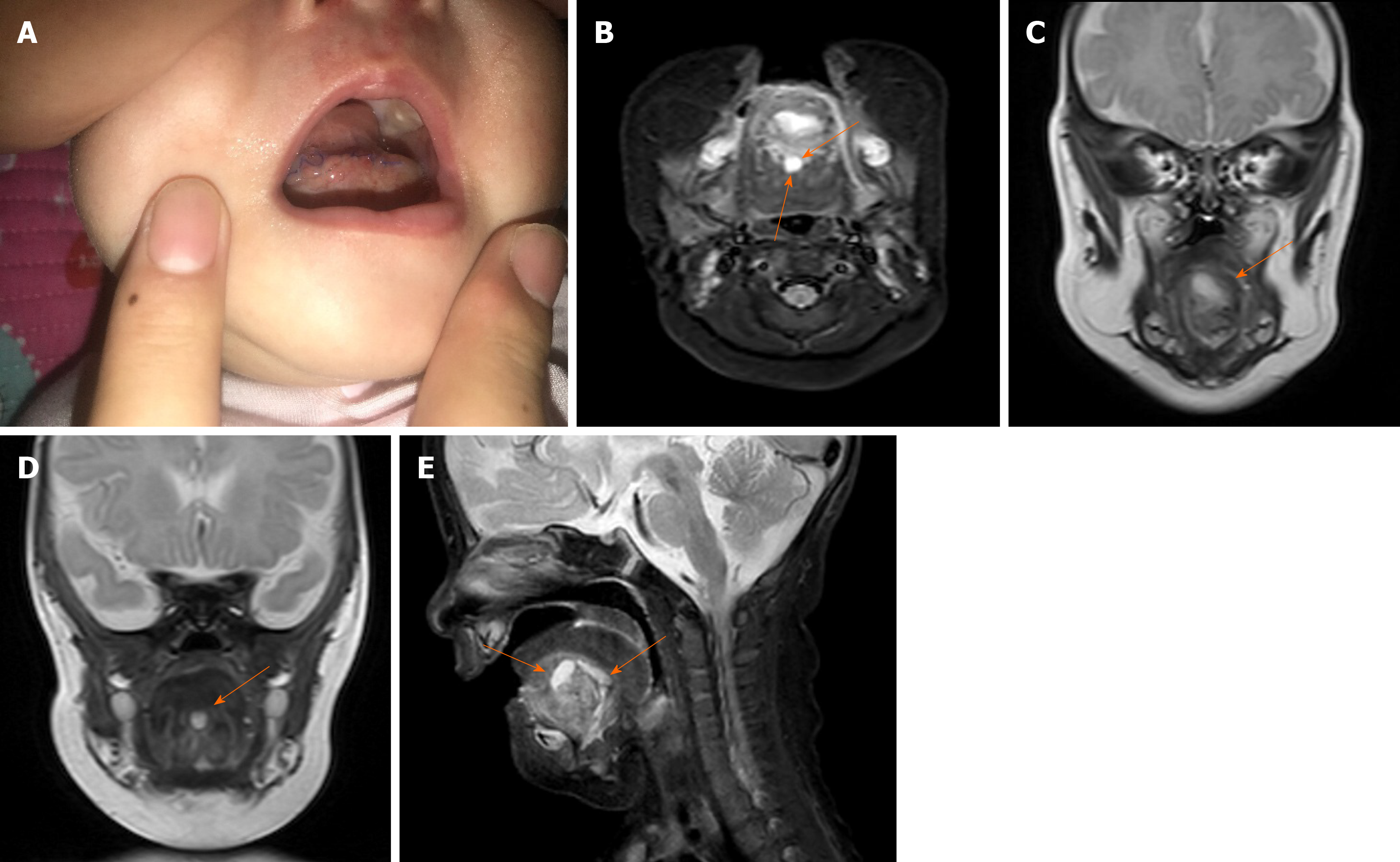
Figure 3 Infant after first surgery (day 3).
A: Slight edema at the floor of the mouth incision; B: Postoperative magnetic resonance imaging showing two cystic lesions (orange arrows) under tongue (0.8 cm × 1.0 cm × 0.5 cm and 1.2 cm × 0.5 cm × 0.5 cm); C and D: View of each lesion in coronal plane (orange arrow); E: Both lesions in sagittal plane (orange arrows).
- Citation: Liu NN, Zhang XY, Tang YY, Wang ZM. Two sequential surgeries in infant with multiple floor of the mouth dermoid cysts: A case report. World J Clin Cases 2020; 8(13): 2885-2892
- URL: https://www.wjgnet.com/2307-8960/full/v8/i13/2885.htm
- DOI: https://dx.doi.org/10.12998/wjcc.v8.i13.2885


