Copyright
©The Author(s) 2020.
World J Clin Cases. Jul 6, 2020; 8(13): 2885-2892
Published online Jul 6, 2020. doi: 10.12998/wjcc.v8.i13.2885
Published online Jul 6, 2020. doi: 10.12998/wjcc.v8.i13.2885
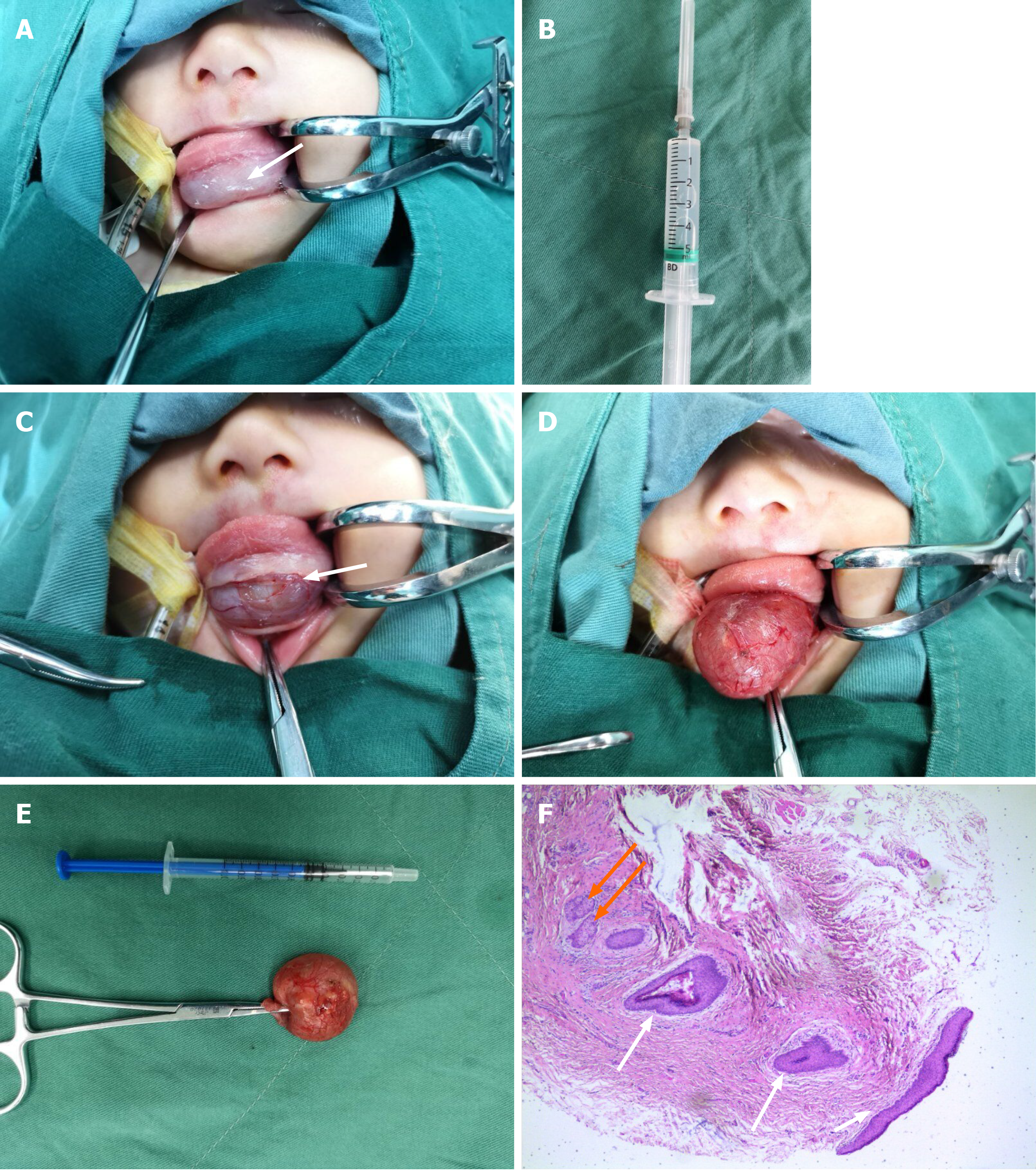
Figure 2 Infant during first surgery.
A: Contracted lesion and lowered tongue after clear fluid (10 mL) aspirated, enabling oral intubation (white arrow at shrunken mass); B: Clear fluid withdrawn; C: Horizontal incision made; D: Careful blunt dissection of cyst from surrounding tissues; E: Resected cyst, roughly 2.0 cm × 2.0 cm with wall intact; F: Histologic section of cyst wall [note keratinizing stratified squamous epithelial lining (white arrows) and sebaceous glands (orange arrows)].
- Citation: Liu NN, Zhang XY, Tang YY, Wang ZM. Two sequential surgeries in infant with multiple floor of the mouth dermoid cysts: A case report. World J Clin Cases 2020; 8(13): 2885-2892
- URL: https://www.wjgnet.com/2307-8960/full/v8/i13/2885.htm
- DOI: https://dx.doi.org/10.12998/wjcc.v8.i13.2885


