Copyright
©The Author(s) 2020.
World J Clin Cases. Jul 6, 2020; 8(13): 2862-2869
Published online Jul 6, 2020. doi: 10.12998/wjcc.v8.i13.2862
Published online Jul 6, 2020. doi: 10.12998/wjcc.v8.i13.2862
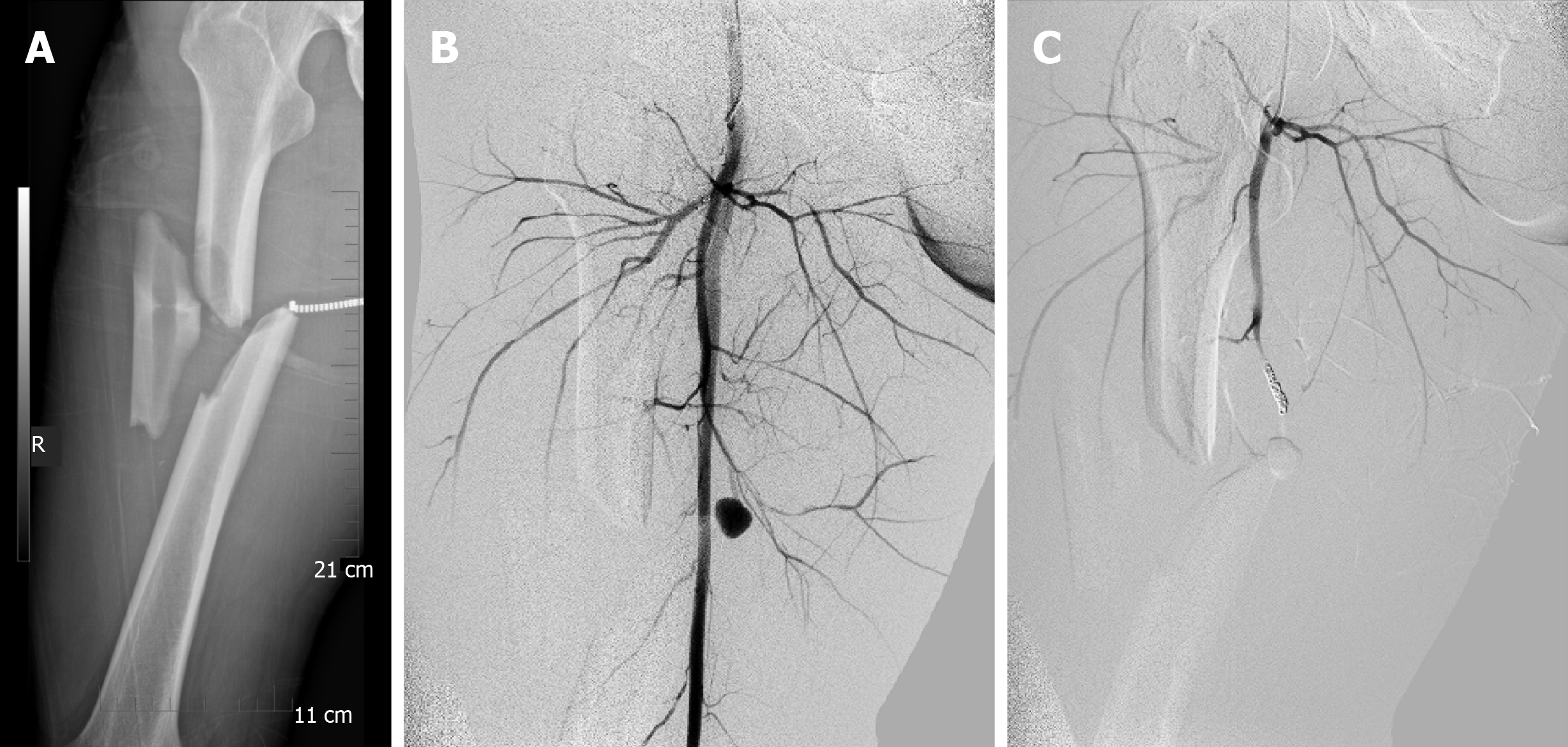
Figure 1 Imaging examinations.
A: Anteroposterior images of right femoral when the patient was delivered to the hospital; B: Computed tomography angiography showed accumulation of the contrast agents at the branch of femoral deep artery, indicating the rupture of the artery; C: A coil was used to embolize the ruptured artery.
- Citation: Ge J, Kong KY, Cheng XQ, Li P, Hu XX, Yang HL, Shen MJ. Missed diagnosis of femoral deep artery rupture after femoral shaft fracture: A case report. World J Clin Cases 2020; 8(13): 2862-2869
- URL: https://www.wjgnet.com/2307-8960/full/v8/i13/2862.htm
- DOI: https://dx.doi.org/10.12998/wjcc.v8.i13.2862


