Copyright
©The Author(s) 2020.
World J Clin Cases. Jul 6, 2020; 8(13): 2841-2848
Published online Jul 6, 2020. doi: 10.12998/wjcc.v8.i13.2841
Published online Jul 6, 2020. doi: 10.12998/wjcc.v8.i13.2841
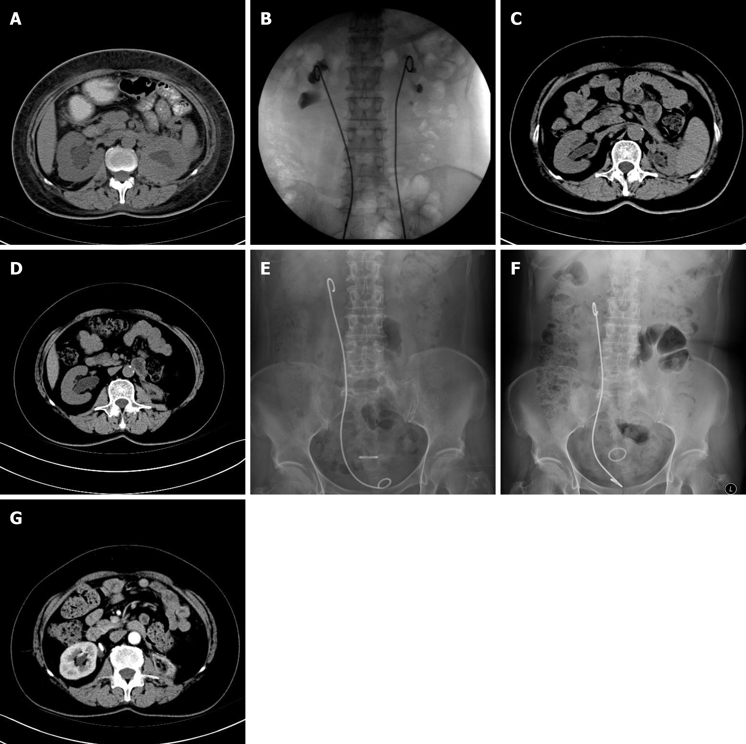
Figure 1 Case 2 received follow-up visits every 3 mo, including ultrasonography.
A: 8 July 2012, the computed tomography (CT) showed bilateral hydronephrosis; B: X-ray guided the placement of metallic stent retrogradely; C: 27 November 2014, the CT scan indicated the left renal atrophy was complete; D: 13 June 2016, the CT showed right kidney severe hydronephrosis. The left kidney was completely atrophied. The patient had renal insufficiency; E: 24 October 2016, the patient had normal renal function, but severe irritative symptoms of bladder. The patient was not tolerant because the distal end of the long metallic stent was in the bladder and exceeded the midline to stimulate the vesical triangle; F: After replacing this stent with a shorter metallic stent (24 cm to substitute for 26 cm), the end of the metallic stent in the bladder did not cross over the midline, and the irritative symptoms of bladder disappeared. The patient tolerated well the shorter metallic stent, and it was replaced every year. Total renal function was normal; G: 8 October 2018, contrast-enhanced CT revealed contrast was well distributed in the right kidney (total renal function). The metallic stent was located in the right ureteropelvic junction. The left kidney was completely atrophied.
- Citation: Gao W, Ou TW, Cui X, Wu JT, Cui B. Metallic ureteral stent in restoring kidney function: Nine case reports. World J Clin Cases 2020; 8(13): 2841-2848
- URL: https://www.wjgnet.com/2307-8960/full/v8/i13/2841.htm
- DOI: https://dx.doi.org/10.12998/wjcc.v8.i13.2841


