Copyright
©The Author(s) 2020.
World J Clin Cases. Jul 6, 2020; 8(13): 2833-2840
Published online Jul 6, 2020. doi: 10.12998/wjcc.v8.i13.2833
Published online Jul 6, 2020. doi: 10.12998/wjcc.v8.i13.2833
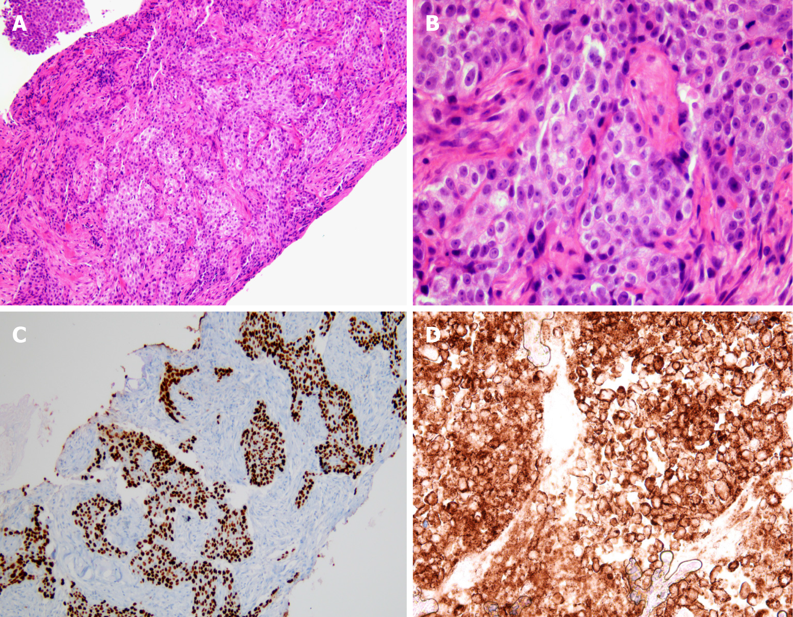
Figure 2 Microscopic examination of the specimen using hematoxylin and eosin staining and immunohistochemistry staining.
A and B: Microscopic examination of the specimen using hematoxylin and eosin staining show keratinized malignant cells (magnification: A, × 100; B, × 400); C: Immunohistochemically, neoplastic cells are positive for p63 (C, × 100); D: The Programmed death-ligand 1 tumor proportion score was ≥ 50% (× 400; PD-L1 IHC 22C3 pharmDx™ Kit, DAKO, Denmark).
- Citation: Kim HB, Park SG, Hong R, Kang SH, Na YS. Acute myelomonocytic leukemia during pembrolizumab treatment for non-small cell lung cancer: A case report. World J Clin Cases 2020; 8(13): 2833-2840
- URL: https://www.wjgnet.com/2307-8960/full/v8/i13/2833.htm
- DOI: https://dx.doi.org/10.12998/wjcc.v8.i13.2833


