Copyright
©The Author(s) 2020.
World J Clin Cases. Jul 6, 2020; 8(13): 2833-2840
Published online Jul 6, 2020. doi: 10.12998/wjcc.v8.i13.2833
Published online Jul 6, 2020. doi: 10.12998/wjcc.v8.i13.2833
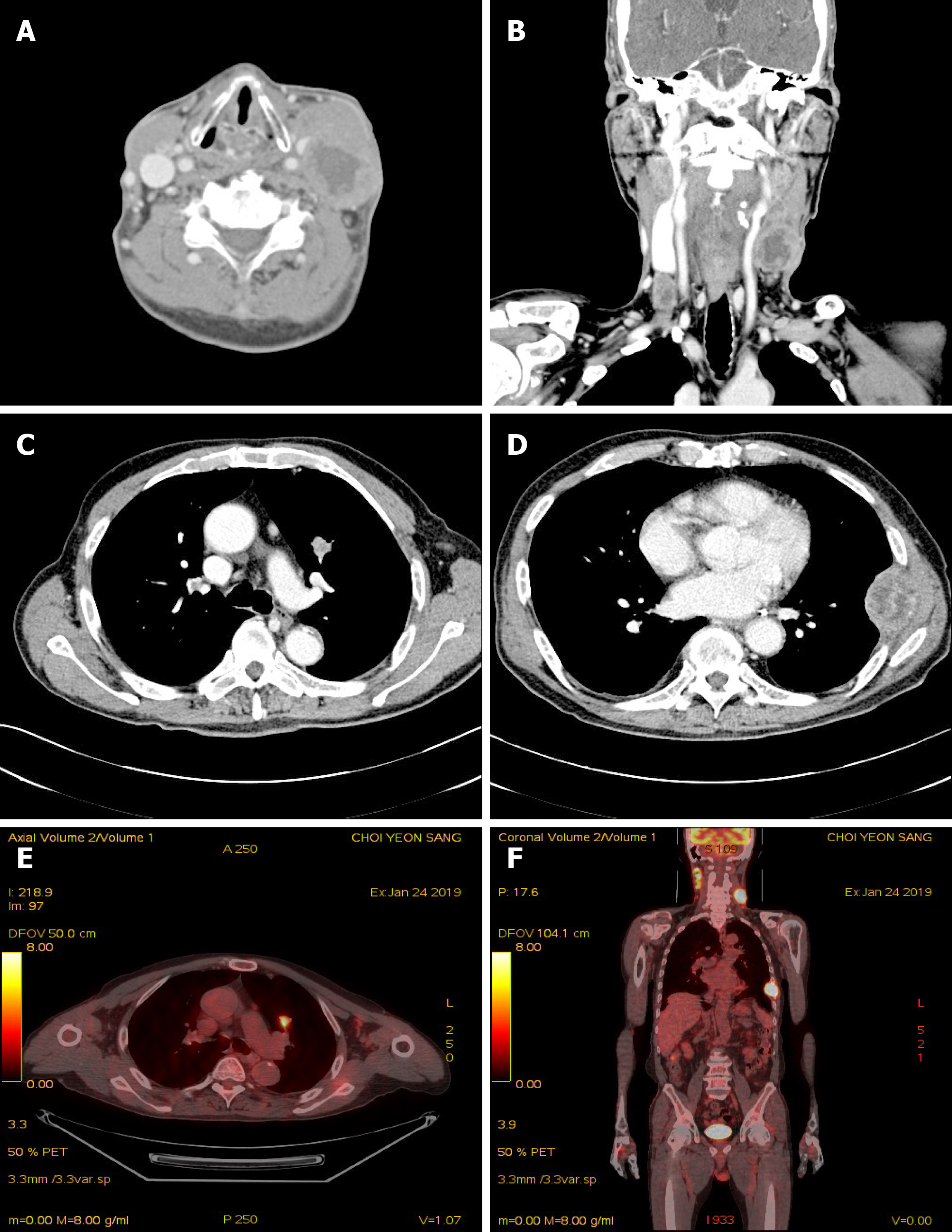
Figure 1 Contrast-enhanced neck computed tomography, contrast-enhanced chest computed tomography, and 18F-fluorodeoxyglucose positron emission/computed tomography.
A: 41 mm × 64 mm sized bulky necrotizing lymphadenopathy in left cervical level V; B: Multiple various sized necrotizing lymphadenopathy on both cervical level II to V; C: Contrast-enhanced chest computed tomography shows 2 cm sized heterogeneous enhanced nodule in anterior segment of left upper lobe (LUL); D: Large periosseous mass formation involving lateral arc of left 7th rib; E: 18F-fluorodeoxyglucose positron emission/computed tomography showed a hypermetabolic nodule in LUL; F: Multiple enlarged hypermetabolic mass on both cervical level II to IV, and hypermetabolic mass on left 7th rib.
- Citation: Kim HB, Park SG, Hong R, Kang SH, Na YS. Acute myelomonocytic leukemia during pembrolizumab treatment for non-small cell lung cancer: A case report. World J Clin Cases 2020; 8(13): 2833-2840
- URL: https://www.wjgnet.com/2307-8960/full/v8/i13/2833.htm
- DOI: https://dx.doi.org/10.12998/wjcc.v8.i13.2833


