Copyright
©The Author(s) 2020.
World J Clin Cases. Jun 26, 2020; 8(12): 2662-2666
Published online Jun 26, 2020. doi: 10.12998/wjcc.v8.i12.2662
Published online Jun 26, 2020. doi: 10.12998/wjcc.v8.i12.2662
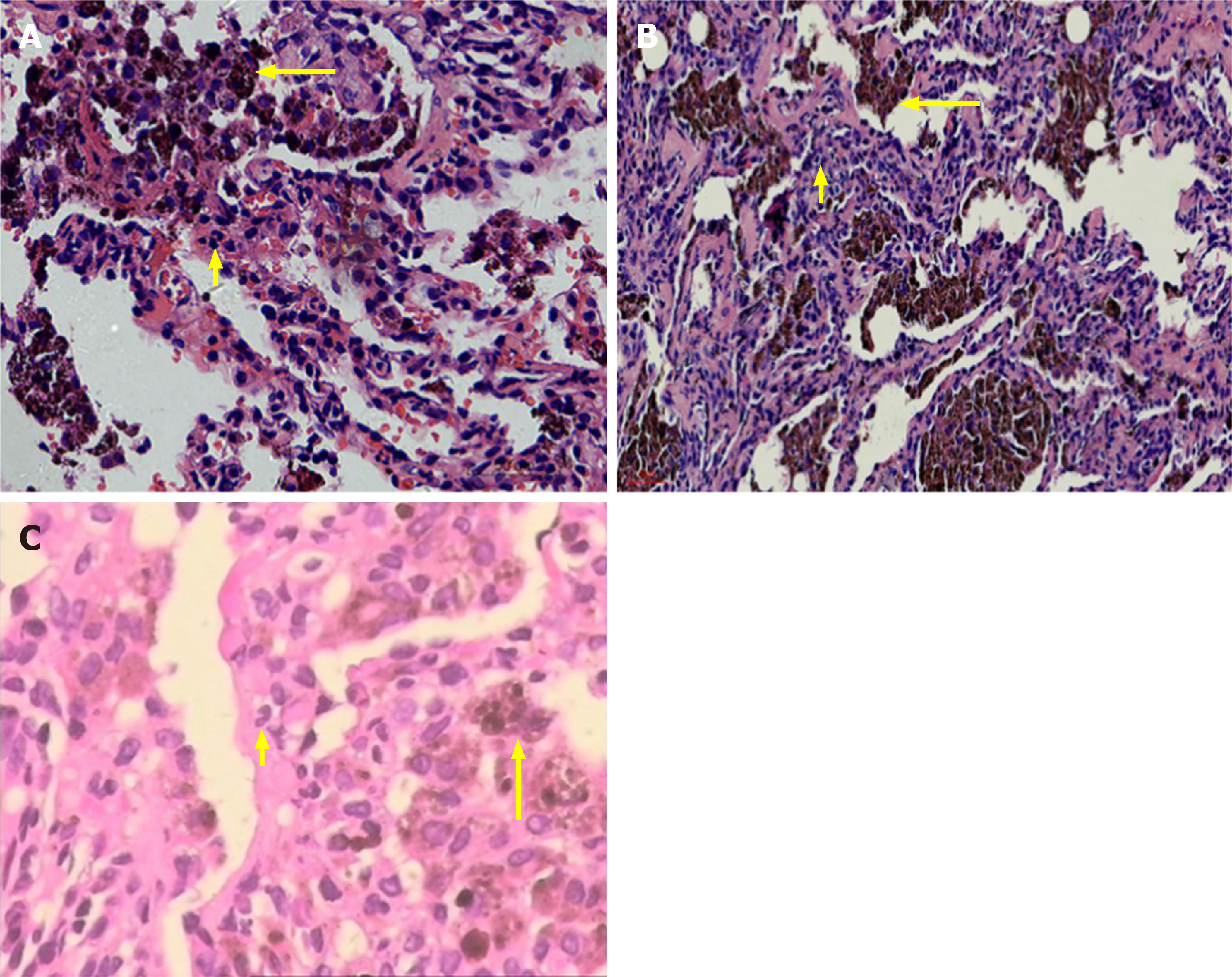
Figure 2 Hematoxylin and eosin staining.
A: Aggregation of hemosiderin-laden macrophage cells (HLMs) within the alveolar compartment (long arrow), alveolar interval are mild fibrous thickening and neutrophil infiltrate (short arrow) (400 ×); B: Acute extravasation of fibrin and red blood cells, HLM deposits in the alveolar cavities (long arrow) and neutrophil infiltrates (short arrow) (400 ×); C: Partial lung tissue fibrosis and HLM deposits were observed in the alveolar cavities (long arrow) and neutrophil infiltrates (short arrow) (400 ×).
- Citation: Xie J, Zhao YY, Liu J, Nong GM. Diffuse alveolar hemorrhage with histopathologic manifestations of pulmonary capillaritis: Three case reports. World J Clin Cases 2020; 8(12): 2662-2666
- URL: https://www.wjgnet.com/2307-8960/full/v8/i12/2662.htm
- DOI: https://dx.doi.org/10.12998/wjcc.v8.i12.2662


