Copyright
©The Author(s) 2020.
World J Clin Cases. Jun 26, 2020; 8(12): 2647-2654
Published online Jun 26, 2020. doi: 10.12998/wjcc.v8.i12.2647
Published online Jun 26, 2020. doi: 10.12998/wjcc.v8.i12.2647
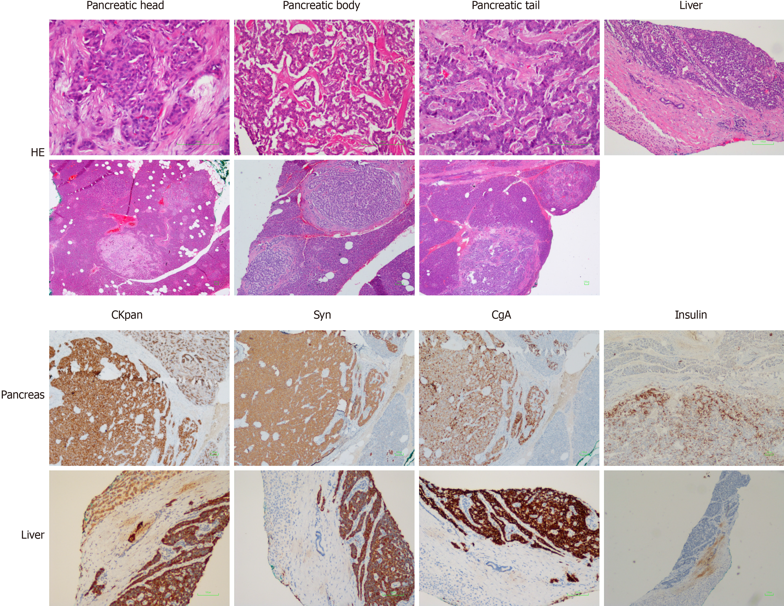
Figure 4 Pathological examination.
Hematoxylin & eosin staining showed (magnification, 40 ×; 100 ×) multiple nodules next to the pancreatic tumor, in the pancreatic tissue around the pancreatic body, and next to the pancreatic tail. Immunohistochemistry showed (magnification, 40 ×; 100 ×) CKpan (+), synaptophysin (+), chromogranin A (+), partial CD56 (+), p53 (-), partial PGP9.5 (+), partial SSTR2 (+), CD10 (-), partial vimentin (+) in pancreas tissues, and CKpan (+), Syn (+), CgA (+), partial CD56 (+), p53 (-), and PGP9.5 (+) in liver tissues. Bar, 100 μm. Syn: Synaptophysin; CgA: Chromogranin A.
- Citation: Ma CH, Guo HB, Pan XY, Zhang WX. Comprehensive treatment of rare multiple endocrine neoplasia type 1: A case report. World J Clin Cases 2020; 8(12): 2647-2654
- URL: https://www.wjgnet.com/2307-8960/full/v8/i12/2647.htm
- DOI: https://dx.doi.org/10.12998/wjcc.v8.i12.2647


