Copyright
©The Author(s) 2020.
World J Clin Cases. Jun 26, 2020; 8(12): 2647-2654
Published online Jun 26, 2020. doi: 10.12998/wjcc.v8.i12.2647
Published online Jun 26, 2020. doi: 10.12998/wjcc.v8.i12.2647
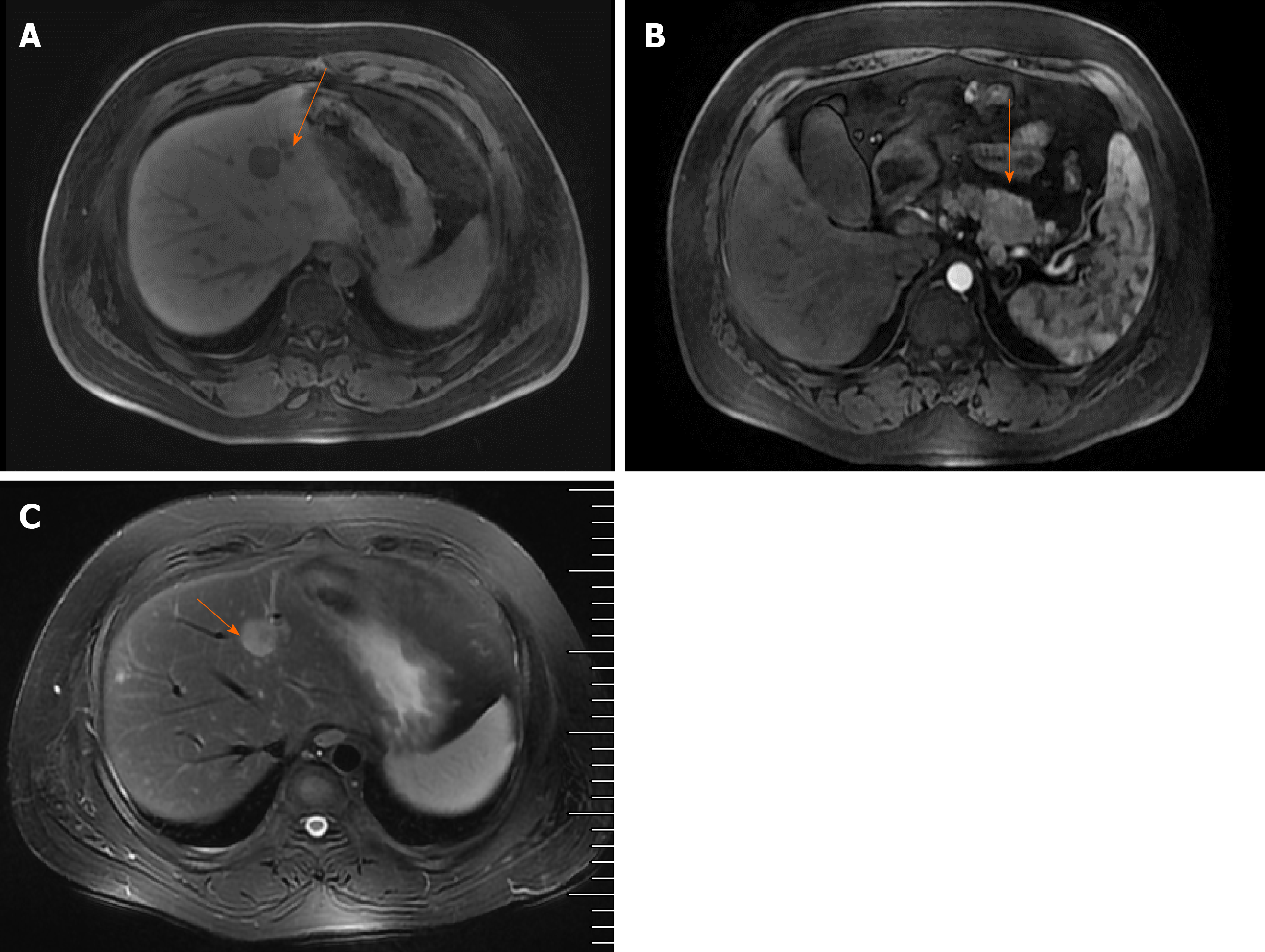
Figure 2 resonance imaging (MRI) examination revealed multiple abnormal signals in the liver segment II, segment VIII, and the junction area of liver segment II and IV as well as occupied lesions in the tail of the pancreatic body; B: Enhanced MRI revealed occupied lesions in the body and tail of the pancreas; C: Enhanced MRI revealed that the liver segment IV was occupied, suggesting the possibility of angiomyolipoma.
There were also small cysts in segments II and VIII of the liver and occupied lesions in the tail of the pancreatic body.
- Citation: Ma CH, Guo HB, Pan XY, Zhang WX. Comprehensive treatment of rare multiple endocrine neoplasia type 1: A case report. World J Clin Cases 2020; 8(12): 2647-2654
- URL: https://www.wjgnet.com/2307-8960/full/v8/i12/2647.htm
- DOI: https://dx.doi.org/10.12998/wjcc.v8.i12.2647


