Copyright
©The Author(s) 2020.
World J Clin Cases. Jun 26, 2020; 8(12): 2603-2609
Published online Jun 26, 2020. doi: 10.12998/wjcc.v8.i12.2603
Published online Jun 26, 2020. doi: 10.12998/wjcc.v8.i12.2603
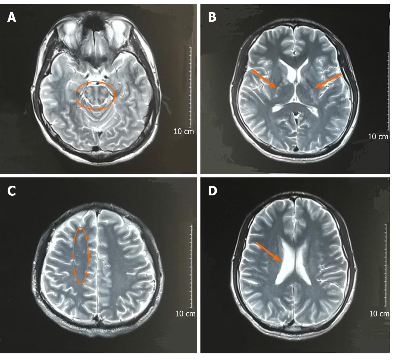
Figure 1 Brain magnetic resonance imaging scans 1 mo before admission.
A: T2 weighted image shows high signal of the brain stem (orange circle); B: T2 weighted image shows high signal of bilateral internal capsules (orange arrows); C: T2 weighted image shows high signal of the right of frontal lobe (orange circle); D: T2 weighted image shows that the right ventricle is enlarged (orange arrow).
- Citation: Li XY, Shi ZH, Guan YL, Ji Y. Anti-N-methyl-D-aspartate-receptor antibody encephalitis combined with syphilis: A case report. World J Clin Cases 2020; 8(12): 2603-2609
- URL: https://www.wjgnet.com/2307-8960/full/v8/i12/2603.htm
- DOI: https://dx.doi.org/10.12998/wjcc.v8.i12.2603


