Copyright
©The Author(s) 2020.
World J Clin Cases. Jun 6, 2020; 8(11): 2399-2405
Published online Jun 6, 2020. doi: 10.12998/wjcc.v8.i11.2399
Published online Jun 6, 2020. doi: 10.12998/wjcc.v8.i11.2399
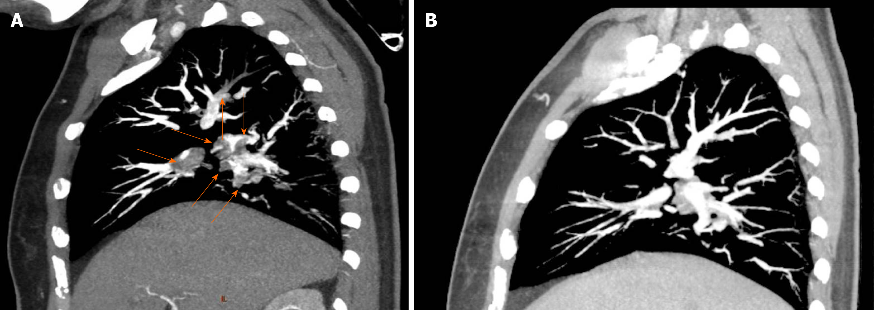
Figure 1 Representative computed tomography angiography evidence of pulmonary embolism and follow-up recovery.
A: Four years before this admission, the patient was diagnosed with pulmonary embolism. This is one representative computed tomography angiography image of the pulmonary embolism. Red arrows indicate the thrombi inside the pulmonary arteries; B: Following 6 mo of treatment after the diagnosis of pulmonary embolism, follow-up pulmonary computed tomography angiography showed obvious thrombus regression.
- Citation: Du BB, Wang XT, Tong YL, Liu K, Li PP, Li XD, Yang P, Wang Y. Optical coherence tomography guided treatment avoids stenting in an antiphospholipid syndrome patient: A case report. World J Clin Cases 2020; 8(11): 2399-2405
- URL: https://www.wjgnet.com/2307-8960/full/v8/i11/2399.htm
- DOI: https://dx.doi.org/10.12998/wjcc.v8.i11.2399


