Copyright
©The Author(s) 2020.
World J Clin Cases. Jun 6, 2020; 8(11): 2364-2373
Published online Jun 6, 2020. doi: 10.12998/wjcc.v8.i11.2364
Published online Jun 6, 2020. doi: 10.12998/wjcc.v8.i11.2364
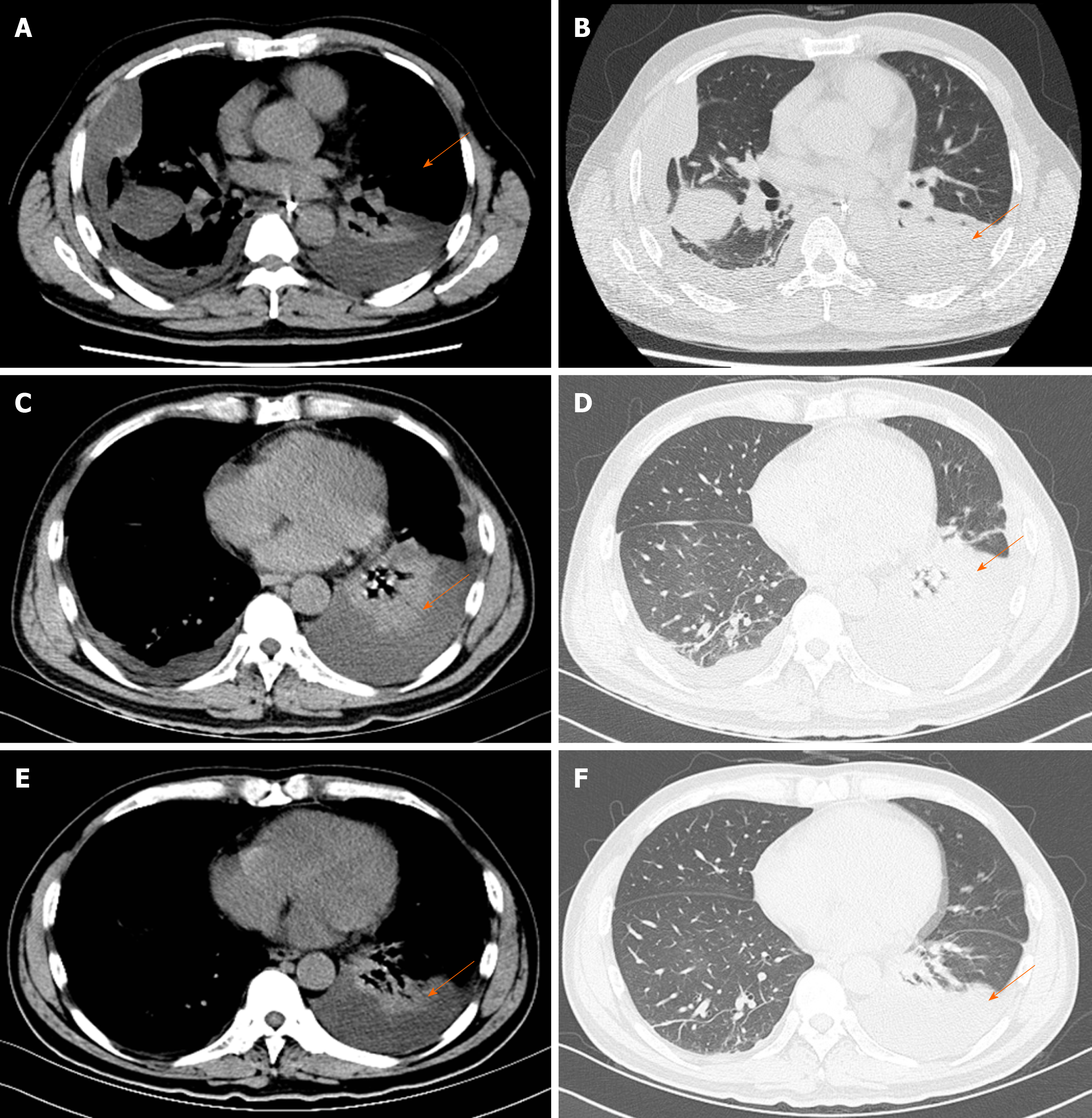
Figure 3 Images of thoracic computed tomography.
A and B: On the fourth day, thoracic computed tomography showed bilateral pleural effusion with partially encapsulated effusion. The lungs also showed scattered linear and lamellar high-density shadows, which were considered to be due to an infectious disease; C and D: Thoracic computed tomography indicated that encapsulated effusion was gradually absorbed but the left lung showed segmental atelectasis on day 13; E and F: Bilateral pleural effusion completely disappeared and segmental atelectasis was recovered on day 23.
- Citation: Han CQ, Xie XR, Zhang Q, Ding Z, Hou XH. Hemophagocytic syndrome as a complication of acute pancreatitis: A case report. World J Clin Cases 2020; 8(11): 2364-2373
- URL: https://www.wjgnet.com/2307-8960/full/v8/i11/2364.htm
- DOI: https://dx.doi.org/10.12998/wjcc.v8.i11.2364


