Copyright
©The Author(s) 2020.
World J Clin Cases. Jun 6, 2020; 8(11): 2350-2358
Published online Jun 6, 2020. doi: 10.12998/wjcc.v8.i11.2350
Published online Jun 6, 2020. doi: 10.12998/wjcc.v8.i11.2350
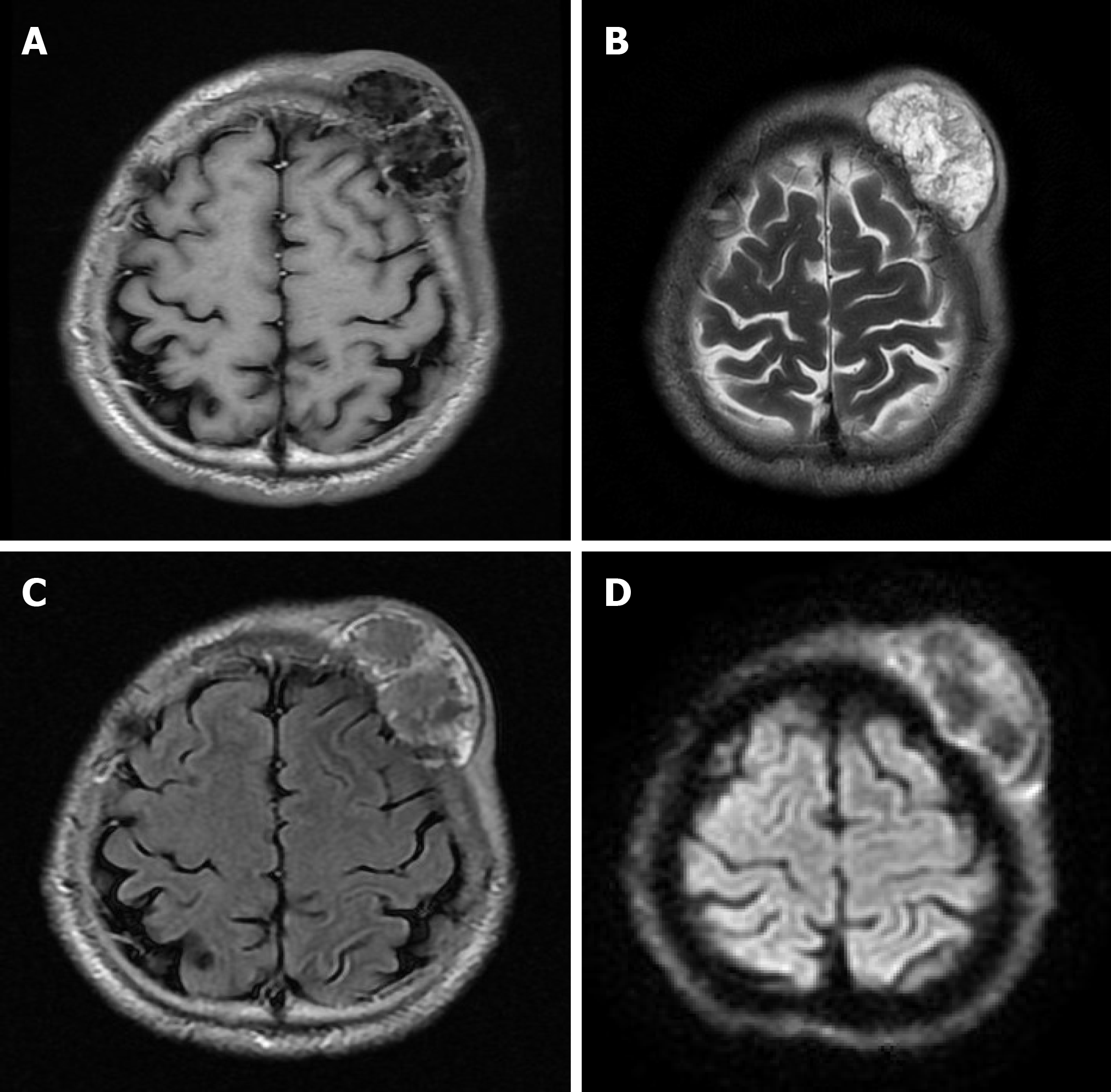
Figure 2 The magnetic resonance imaging revealed an irregular lesion, approximately 3.
55 cm × 6.34 cm in size, in the left frontal region with clear boundaries and visible separation. The adjacent skull was damaged and the dura mater was involved. A: T1 showed complex signal with a dramatic low signal; B: T2 showed complex signal with a dramatic high signal; C: T2 FLAIR showed high marginal and low central signals; D: DWI showed high marginal and low central signals.
- Citation: Ke XT, Yu XF, Liu JY, Huang F, Chen MG, Lai QQ. Myxofibrosarcoma of the scalp with difficult preoperative diagnosis: A case report and review of the literature. World J Clin Cases 2020; 8(11): 2350-2358
- URL: https://www.wjgnet.com/2307-8960/full/v8/i11/2350.htm
- DOI: https://dx.doi.org/10.12998/wjcc.v8.i11.2350


