Copyright
©The Author(s) 2020.
World J Clin Cases. Jun 6, 2020; 8(11): 2345-2349
Published online Jun 6, 2020. doi: 10.12998/wjcc.v8.i11.2345
Published online Jun 6, 2020. doi: 10.12998/wjcc.v8.i11.2345
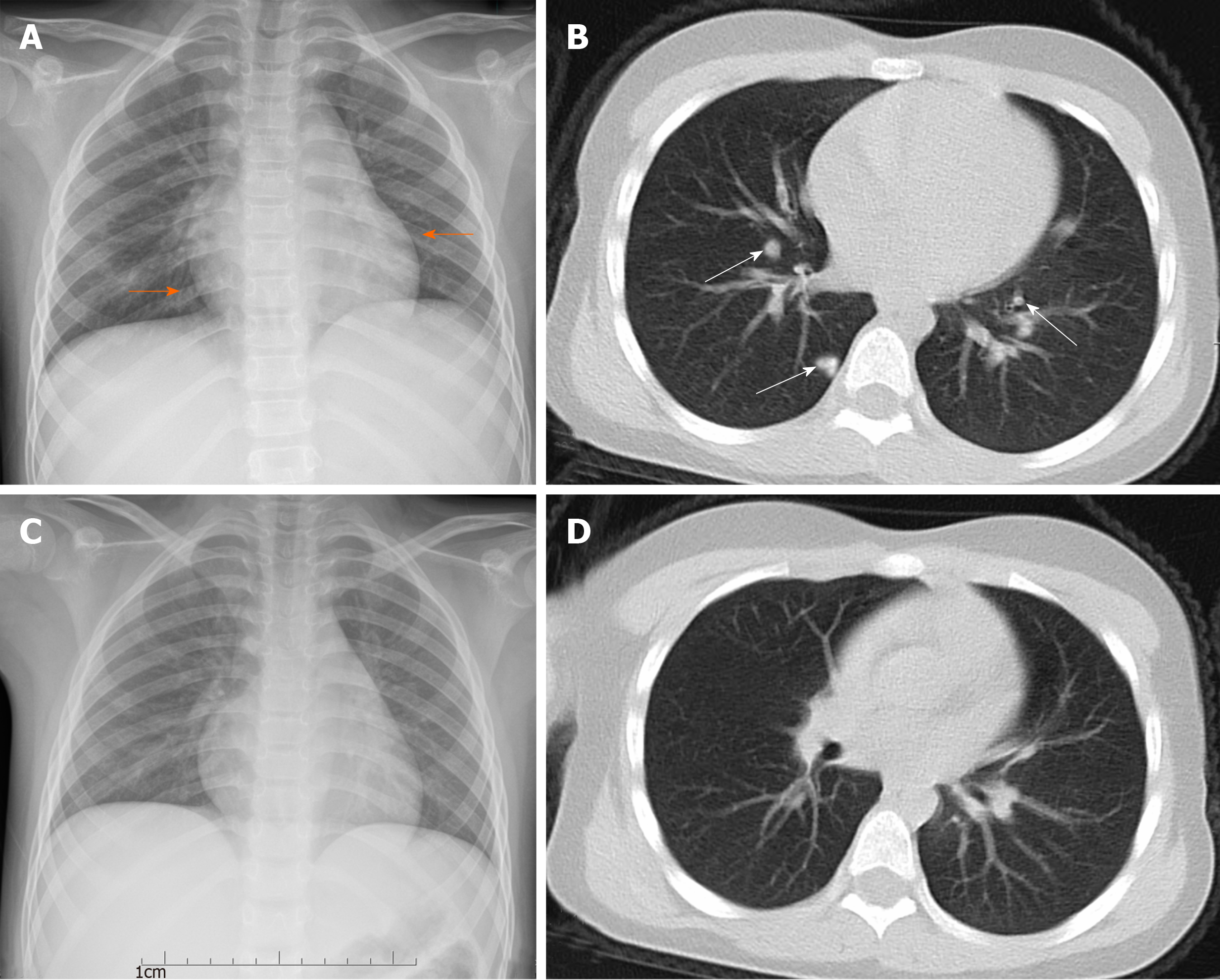
Figure 1 Seven-year-old girl with coronavirus disease 2019 pneumonia.
The posteroanterior chest radiograph and pectoral computed tomography (CT) scan were carried out in Huzhou Central Hospital on the first day of admission (1 d after fever) (A and B) and 3 d after antiviral therapy (C and D). A: Chest radiograph showed a patchy increase in the opacity of the left lower lung area located in the retrocardiac region (orange arrows); B: CT scan showed multiple patchy consolidations in both upper lobes with a ground-glass opacity halo and nodular lesions (white arrows); C: Chest radiograph showed the newly discovered unclear patchy area increase in the opacity of the right upper lung area; D: Coronal reformation CT showed rapid focal absorption in the area of pulmonary infection after 3 d of antiviral therapy.
- Citation: Chen X, Zou XJ, Xu Z. Serial computed tomographic findings and specific clinical features of pediatric COVID-19 pneumonia: A case report. World J Clin Cases 2020; 8(11): 2345-2349
- URL: https://www.wjgnet.com/2307-8960/full/v8/i11/2345.htm
- DOI: https://dx.doi.org/10.12998/wjcc.v8.i11.2345


