Copyright
©The Author(s) 2020.
World J Clin Cases. Jun 6, 2020; 8(11): 2219-2226
Published online Jun 6, 2020. doi: 10.12998/wjcc.v8.i11.2219
Published online Jun 6, 2020. doi: 10.12998/wjcc.v8.i11.2219
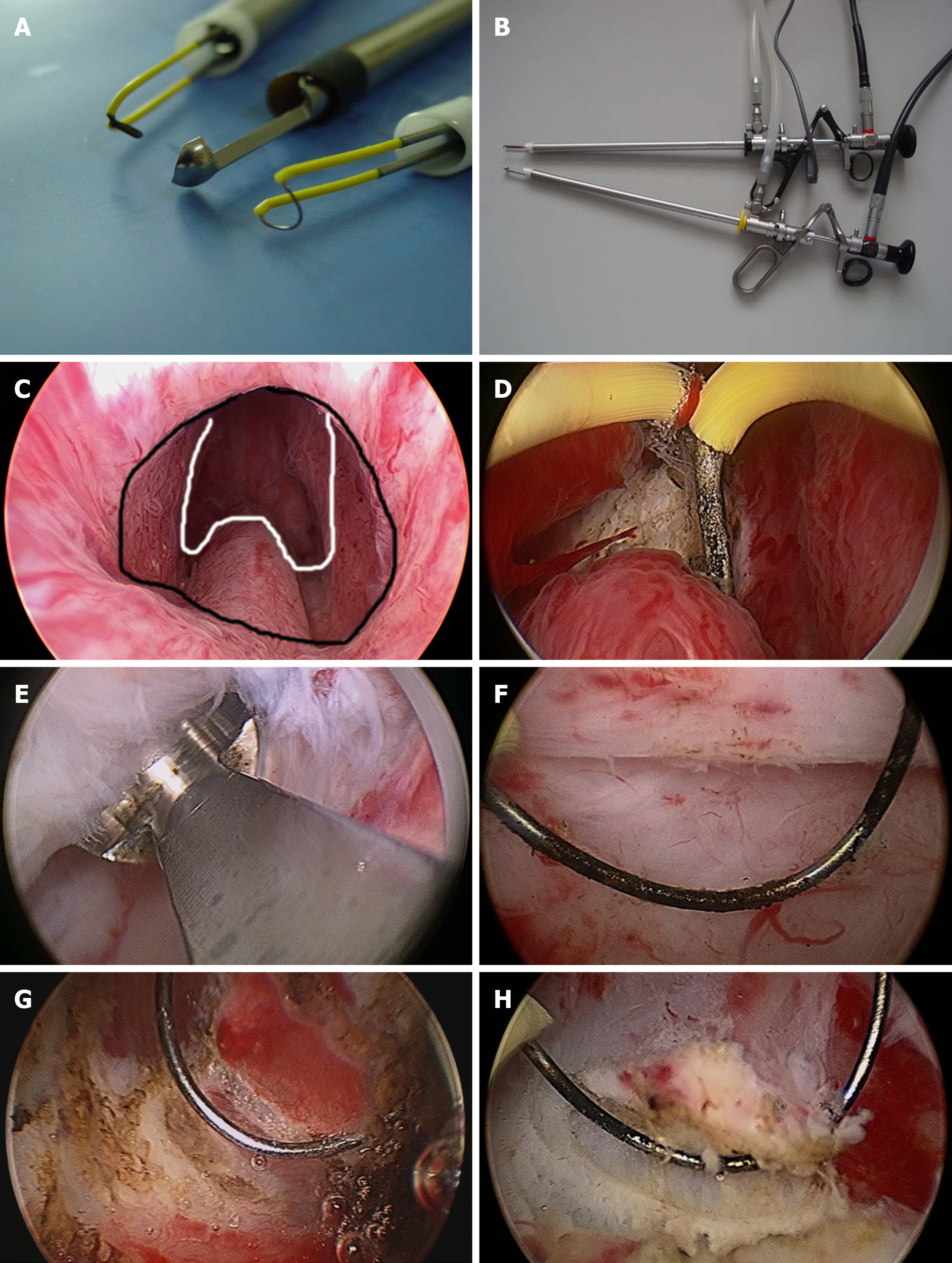
Figure 1 Instruments and procedure of Hiraoka's transurethral detachment of prostate combined with biopsy of the peripheral zone.
A: Surgical instruments (from top-to-bottom): Needle electrode, Hiraoka's Prostate Detaching Blade (the tip of the detaching blade is a shallow, sharp-edged scoop with two longitudinal apertures to enhance the flow of irrigation fluid) and a cutting loop electrode; B: Hiraoka's Prostate Detaching Blade can be connected with a 26 Fr continuous irrigation sheath and resectoscope; C: Nesbit sign: the white solid line indicates the distal part of the prostate adenoma; the black solid line indicates the proximal part of the urethral external sphincter; D: A needle electrode was used to incise the urethral mucosa in a circular incision along the marker, then Hiraoka's Prostate Detaching Blade was used to locate and enter the surgical capsule plane at the 5-7 o'clock position of the circular incision; E: The hyperplastic prostate gland is white with a dense fibrous structure, and the surgical capsule is spongy with soft and transverse vessels when visualized with a resectoscope; F: There was little bleeding during the detaching process, which generally does not affect the visual field. If necessary, we could use a cutting loop electrode to stop bleeding; G and H: Biopsy of the lateral peripheral zone with a cutting loop electrode.
- Citation: Pan CY, Wu B, Yao ZC, Zhu XQ, Jiang YZ, Bai S. Role of Hiraoka's transurethral detachment of the prostate combined with biopsy of the peripheral zone during the same session in patients with repeated negative biopsies in the diagnosis of prostate cancer. World J Clin Cases 2020; 8(11): 2219-2226
- URL: https://www.wjgnet.com/2307-8960/full/v8/i11/2219.htm
- DOI: https://dx.doi.org/10.12998/wjcc.v8.i11.2219


