Copyright
©The Author(s) 2020.
World J Clin Cases. May 26, 2020; 8(10): 1995-2000
Published online May 26, 2020. doi: 10.12998/wjcc.v8.i10.1995
Published online May 26, 2020. doi: 10.12998/wjcc.v8.i10.1995
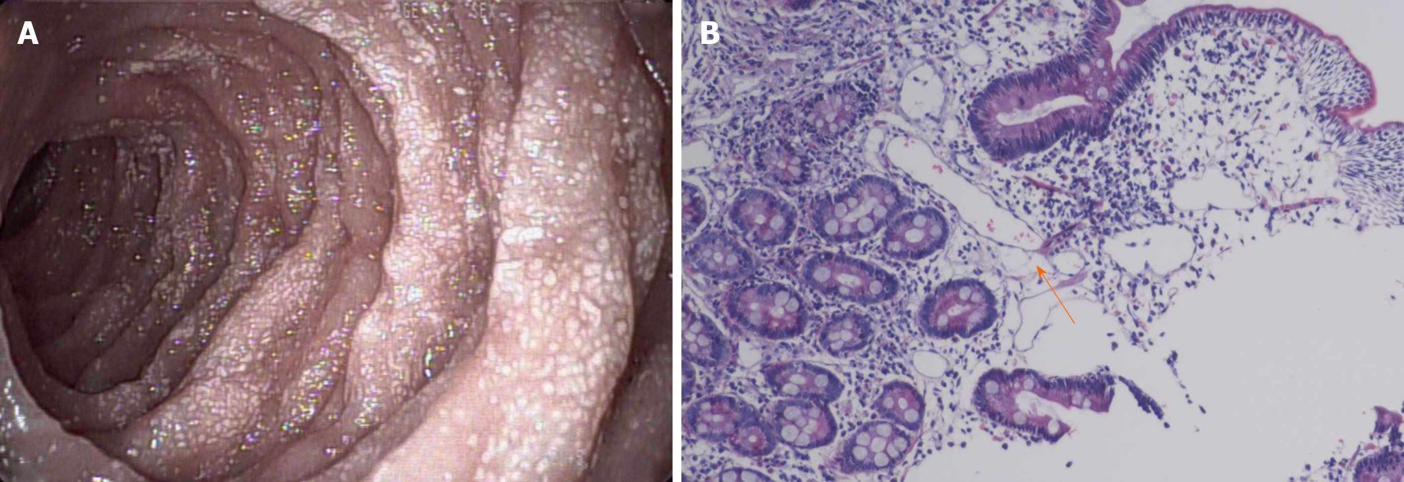
Figure 1 Biopsy findings and histological image.
A: Gastroscopy performed on September 9, 2015 showed that the mucosa of the descending segment and horizontal segment of the duodenum was rough, with dense white spots on the surface; B: Histological image showing dilated lymph vessels (hematoxylin and eosin staining, × 100).
- Citation: Lin WH, Zhang ZH, Wang HL, Ren L, Geng LL. Tuberous sclerosis complex presenting as primary intestinal lymphangiectasia: A case report. World J Clin Cases 2020; 8(10): 1995-2000
- URL: https://www.wjgnet.com/2307-8960/full/v8/i10/1995.htm
- DOI: https://dx.doi.org/10.12998/wjcc.v8.i10.1995


