Copyright
©The Author(s) 2020.
World J Clin Cases. May 26, 2020; 8(10): 1973-1978
Published online May 26, 2020. doi: 10.12998/wjcc.v8.i10.1973
Published online May 26, 2020. doi: 10.12998/wjcc.v8.i10.1973
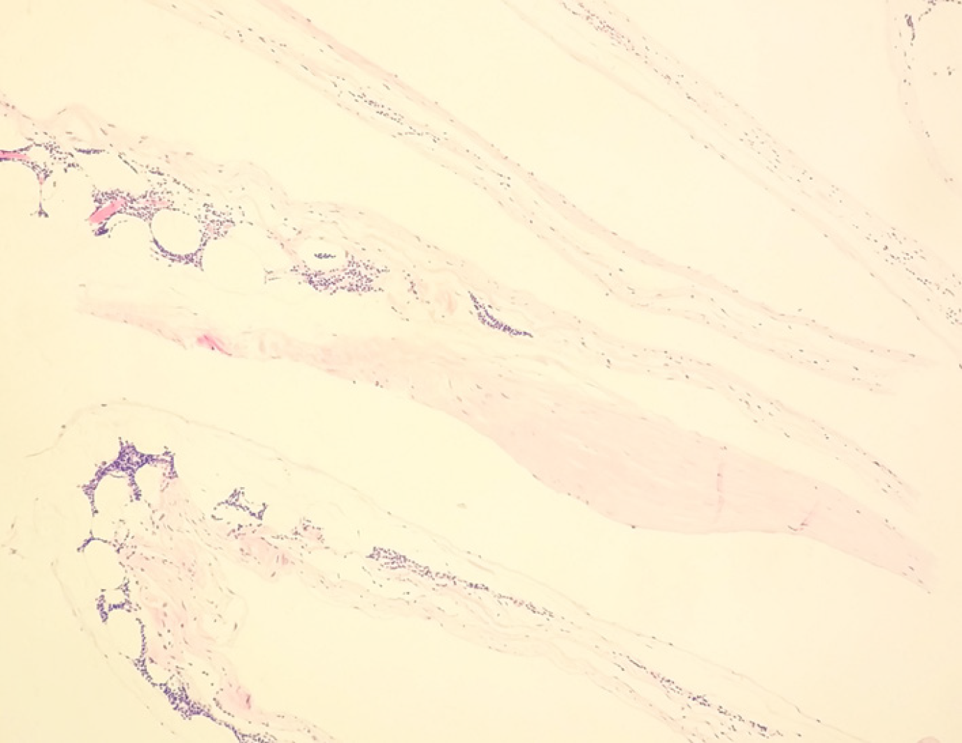
Figure 2 Fibrous cystic wall-like tissue observed under an optical microscope, with proliferated lymphoid tissue inside (HE staining, × 100).
The findings confirmed the lesions as multiple cystic lymphangiomas.
- Citation: Sun MM, Shen J. Positron emission tomography/computed tomography findings of multiple cystic lymphangiomas in an adult: A case report. World J Clin Cases 2020; 8(10): 1973-1978
- URL: https://www.wjgnet.com/2307-8960/full/v8/i10/1973.htm
- DOI: https://dx.doi.org/10.12998/wjcc.v8.i10.1973


