Copyright
©The Author(s) 2020.
World J Clin Cases. May 26, 2020; 8(10): 1950-1957
Published online May 26, 2020. doi: 10.12998/wjcc.v8.i10.1950
Published online May 26, 2020. doi: 10.12998/wjcc.v8.i10.1950
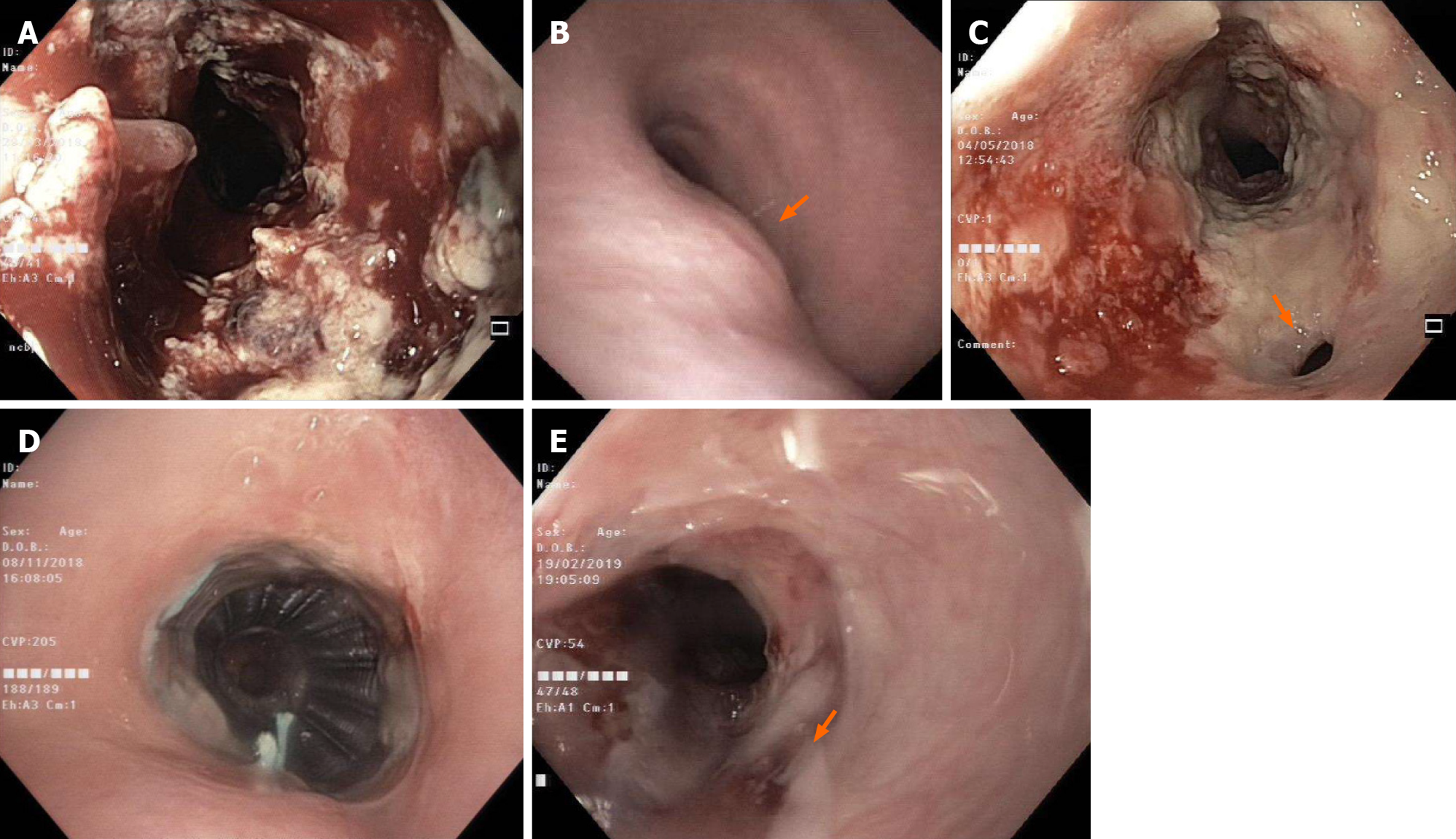
Figure 1 Endoscopic and imaging examinations.
A and B: Esophageal tumor on esophagogastroduodenocopy (A) and airway evaluation on bronchoscopy (B) where an extrinsic compression of trachea (arrow) was observed at initial diagnosis; C: A tracheoesophageal fistula (arrow) seen on esophagogastroduodenocopy after first week of chemoradiotherapy; D: Tracheal stent in place on bronchoscopy at 6-mo post-chemoradiotherapy; E: Complete tumor response with closure of fistula (arrow) evident on esophagogastroduodenocopy at 6-mo post-chemoradiotherapy.
- Citation: Lee CC, Yeo CM, Ng WK, Verma A, Tey JC. T4 cervical esophageal cancer cured with modern chemoradiotherapy: A case report. World J Clin Cases 2020; 8(10): 1950-1957
- URL: https://www.wjgnet.com/2307-8960/full/v8/i10/1950.htm
- DOI: https://dx.doi.org/10.12998/wjcc.v8.i10.1950


