Copyright
©The Author(s) 2020.
World J Clin Cases. May 26, 2020; 8(10): 1887-1896
Published online May 26, 2020. doi: 10.12998/wjcc.v8.i10.1887
Published online May 26, 2020. doi: 10.12998/wjcc.v8.i10.1887
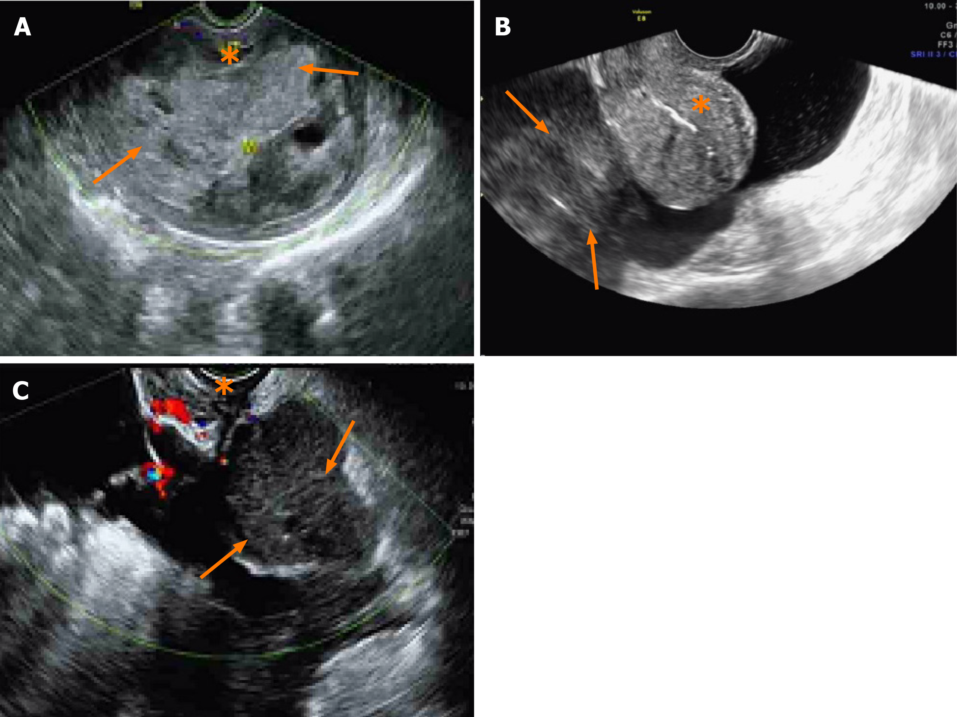
Figure 2 Ultrasound images of uterine leiomyosarcoma and non-uterine leiomyosarcoma.
A: Ultrasound image of a preoperative patient with uterine leiomyosarcoma. Myometrium (asterisk) and intramural lesions of the uterus are shown (arrows); B: Ultrasound image of a preoperative patient with non-uterine leiomyosarcoma. Normal uterus (asterisk) and extrauterine lesions are shown (arrows); C: Ultrasound image of recurrent lesions in leiomyosarcoma. Normal vaginal stump (asterisk) and recurrent pelvic lesions are shown (arrows).
- Citation: Sun Q, Yang X, Zeng Z, Wei X, Li KZ, Xu XY. Outcomes of patients with pelvic leiomyosarcoma treated by surgery and relevant auxiliary diagnosis. World J Clin Cases 2020; 8(10): 1887-1896
- URL: https://www.wjgnet.com/2307-8960/full/v8/i10/1887.htm
- DOI: https://dx.doi.org/10.12998/wjcc.v8.i10.1887


