Copyright
©The Author(s) 2020.
World J Clin Cases. May 26, 2020; 8(10): 1848-1858
Published online May 26, 2020. doi: 10.12998/wjcc.v8.i10.1848
Published online May 26, 2020. doi: 10.12998/wjcc.v8.i10.1848
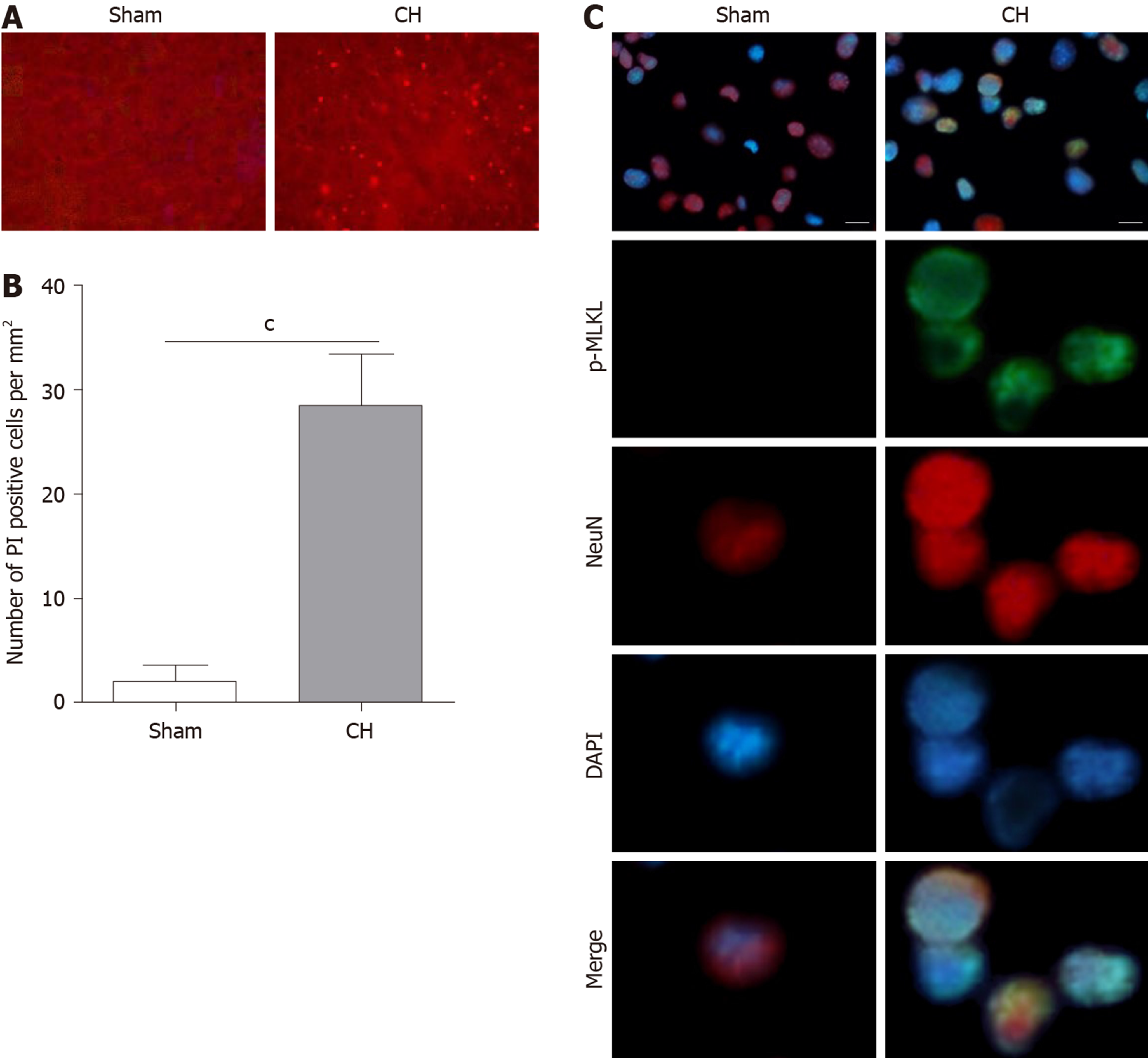
Figure 1 Cerebral hemorrhage induced necroptosis in brain tissue.
A and B: Propidium iodide staining (A) and propidium iodide positive rate (B) in brain tissues of the experimental rats; C: Immunofluorescence assay showed the expression of the necroptotic marker p-MLKL in the experimental rats. Scale bar: 20 μm. cP < 0.001. CH: cerebral hemorrhage; PI: Propidium iodide; MLKL: mixed lineage kinase domain like pseudokinase.
- Citation: Cai GF, Sun ZR, Zhuang Z, Zhou HC, Gao S, Liu K, Shang LL, Jia KP, Wang XZ, Zhao H, Cai GL, Song WL, Xu SN. Cross electro-nape-acupuncture ameliorates cerebral hemorrhage-induced brain damage by inhibiting necroptosis. World J Clin Cases 2020; 8(10): 1848-1858
- URL: https://www.wjgnet.com/2307-8960/full/v8/i10/1848.htm
- DOI: https://dx.doi.org/10.12998/wjcc.v8.i10.1848


