Copyright
©The Author(s) 2020.
World J Clin Cases. May 26, 2020; 8(10): 1832-1847
Published online May 26, 2020. doi: 10.12998/wjcc.v8.i10.1832
Published online May 26, 2020. doi: 10.12998/wjcc.v8.i10.1832
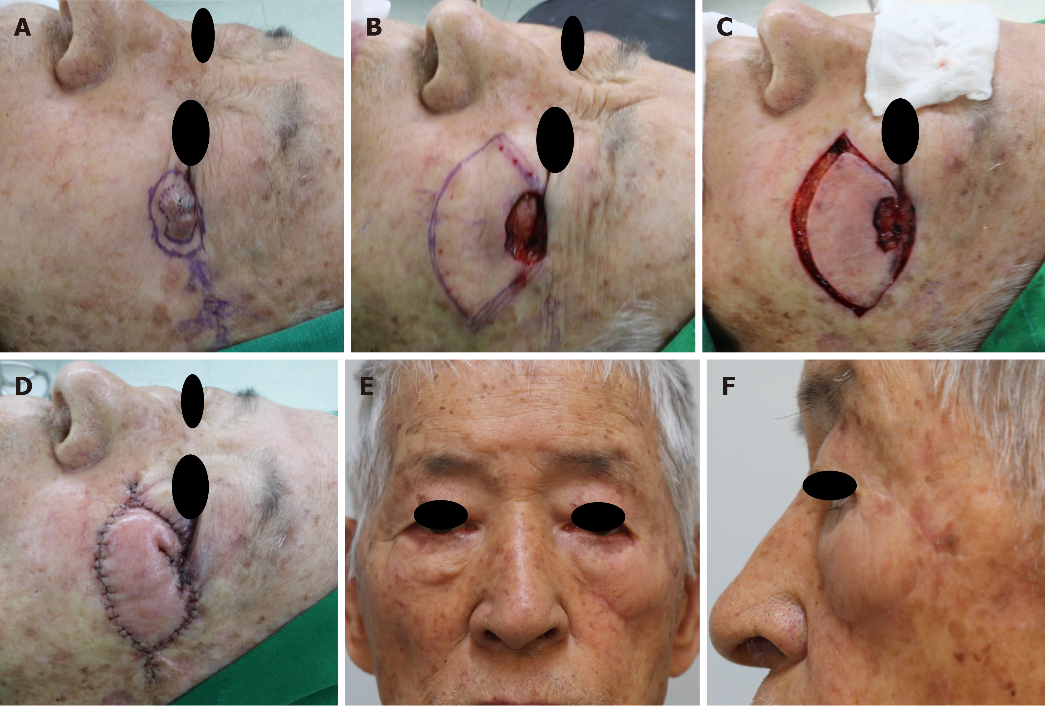
Figure 9 An 86-year-old man was diagnosed with basal cell carcinoma on the left lateral canthal area after a punch biopsy.
A: The lesion was located on the lateral canthal subunit and lower-lid subunit of the left eyelid unit; B: He underwent wide excision of the lesion with a 3-mm safety margin, the final defect was measured to be 2.5 cm × 3 cm; C, D: We covered the defect with a Ω-variant Type IIA keystone design perforator island flap (flap size: 4 cm × 8.5 cm) from the zygomatic subunit and medial subunit of the left cheek unit. Final suture lines were located within and along each facial subunit, and parallel to facial relaxed skin tension lines; E, F: Postoperative clinical photograph after 12 mo.
- Citation: Lim SY, Yoon CS, Lee HG, Kim KN. Keystone design perforator island flap in facial defect reconstruction. World J Clin Cases 2020; 8(10): 1832-1847
- URL: https://www.wjgnet.com/2307-8960/full/v8/i10/1832.htm
- DOI: https://dx.doi.org/10.12998/wjcc.v8.i10.1832


