Copyright
©The Author(s) 2020.
World J Clin Cases. Jan 6, 2020; 8(1): 68-75
Published online Jan 6, 2020. doi: 10.12998/wjcc.v8.i1.68
Published online Jan 6, 2020. doi: 10.12998/wjcc.v8.i1.68
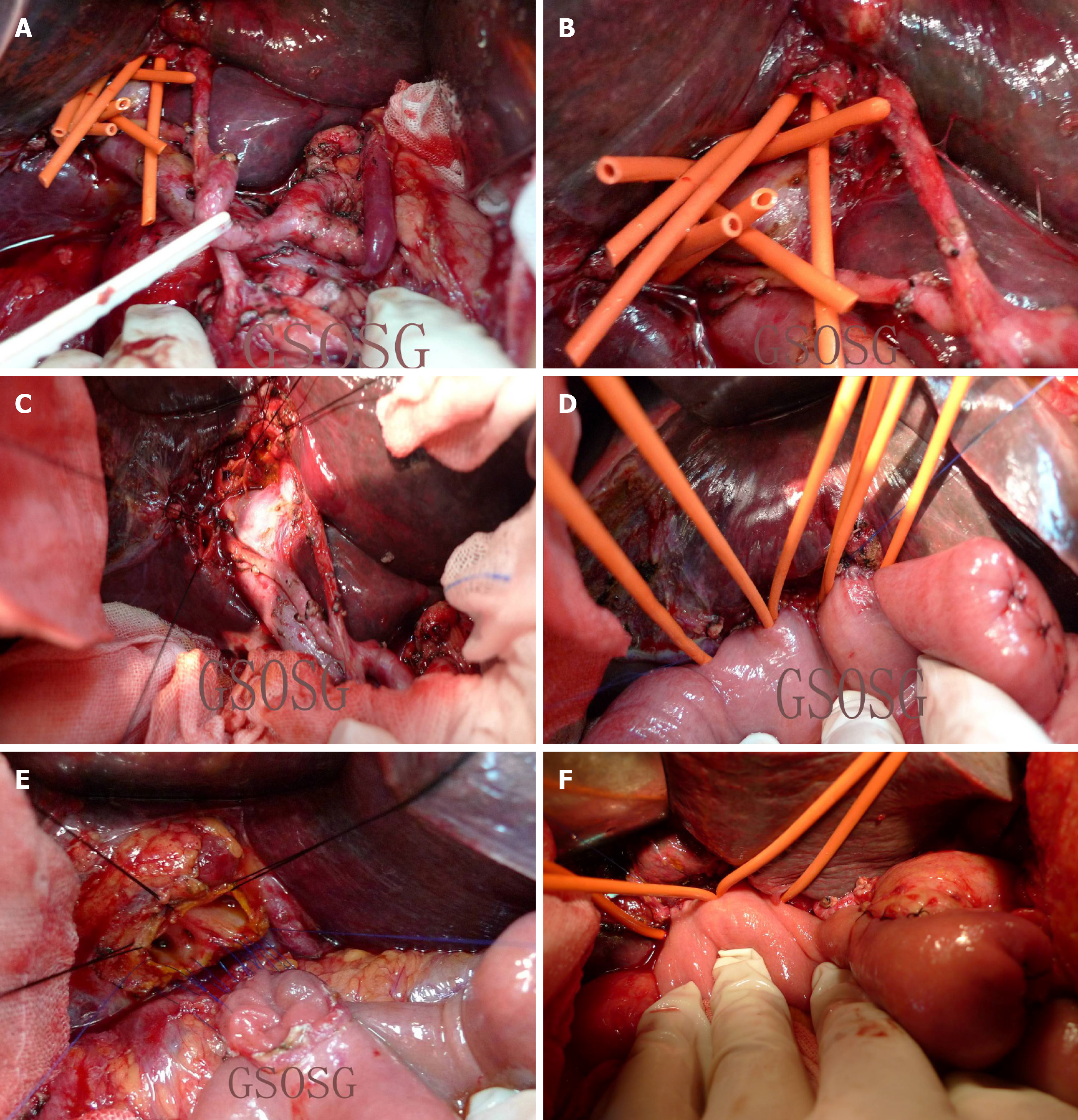
Figure 2 Intraoperative photographs.
A: Complete vascular skeletonization of the hepatoduodenal ligament, including groups 12a, b, c, h, p, 7, 8a, p, 9, and 11d; B: Radical resection and lymphadenectomy of hilar lesion R0 were completed; eight bile duct ends appeared on the remnant hepatic surface of the hilar region, which were hepatic segment ducts, including hepatic segments II, III, and IV; V, VII, and I; and VI and VII; C: Formation of bile duct lake was completed. Hepatic segments II, III, and IV; V, VIII, and I; and VI and VII were used to form a bile duct lake; D: Three of the end-to-side Roux-en-Y hepaticojejunostomy reconstructions by formation of bile duct lake were completed; E: Formation of a bile duct lake was completed using 5-0 Prolene continuous sutures; F: Two of the bile duct lake-forming jejunal end-to-side anastomosis were completed.
- Citation: Yang XJ, Dong XH, Chen SY, Wu B, He Y, Dong BL, Ma BQ, Gao P. Application of multiple Roux-en-Y hepaticojejunostomy reconstruction by formation of bile hilar duct lake in the operation of hilar cholangiocarcinoma. World J Clin Cases 2020; 8(1): 68-75
- URL: https://www.wjgnet.com/2307-8960/full/v8/i1/68.htm
- DOI: https://dx.doi.org/10.12998/wjcc.v8.i1.68


