Copyright
©The Author(s) 2020.
World J Clin Cases. Jan 6, 2020; 8(1): 208-216
Published online Jan 6, 2020. doi: 10.12998/wjcc.v8.i1.208
Published online Jan 6, 2020. doi: 10.12998/wjcc.v8.i1.208
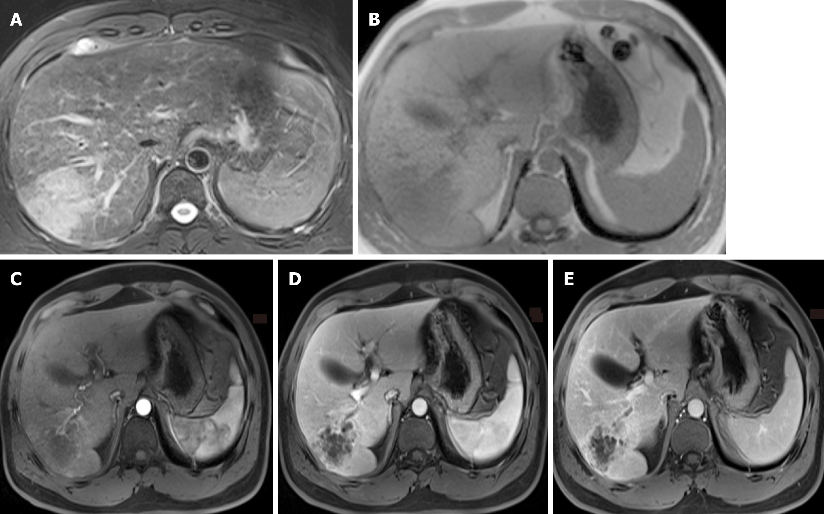
Figure 1 Magnetic resonance imaging results.
A-B: There is an abnormal lesion occupying the right lobe of the liver, hyperintense on non-contrast T2 weighted images and hypointense on non-contrast T1 weighted images; C-E: A honeycomb-like structure and persistent enhancement with slight transient hepatic parenchymal enhancement around it and adjacent proximal bile duct dilatation with enhancement of the wall on contrast-enhanced T1 weighted images were observed.
- Citation: Wang Y, Ming JL, Ren XY, Qiu L, Zhou LJ, Yang SD, Fang XM. Sarcomatoid intrahepatic cholangiocarcinoma mimicking liver abscess: A case report. World J Clin Cases 2020; 8(1): 208-216
- URL: https://www.wjgnet.com/2307-8960/full/v8/i1/208.htm
- DOI: https://dx.doi.org/10.12998/wjcc.v8.i1.208


