Copyright
©The Author(s) 2020.
World J Clin Cases. Jan 6, 2020; 8(1): 179-187
Published online Jan 6, 2020. doi: 10.12998/wjcc.v8.i1.179
Published online Jan 6, 2020. doi: 10.12998/wjcc.v8.i1.179
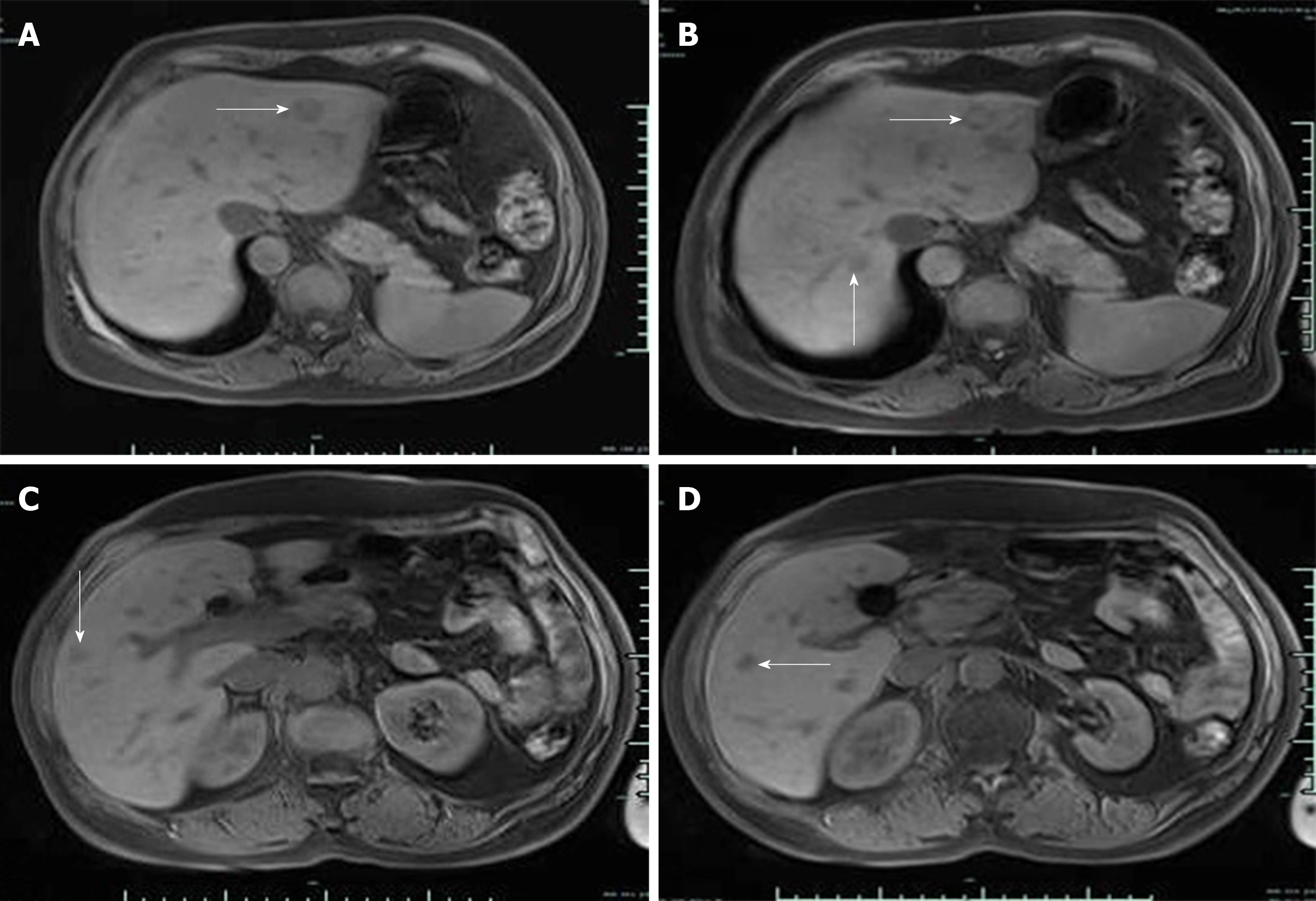
Figure 4 Magnetic resonance images of liver metastases.
A: An oval mass in the left liver before transcatheter arterial chemoembolization with a clear boundary and diameter of about 1.9 cm; B, C, D: Magnetic resonance imaging 3 mo after transcatheter arterial chemoembolization revealed no significant reduction in the original metastatic tumor in the left liver and appearance of a new metastatic tumor in the right liver (arrow).
- Citation: Cai HJ, Wang H, Cao N, Huang B, Kong FL, Lu LR, Huang YY, Wang W. Calcitonin-negative neuroendocrine tumor of the thyroid with metastasis to liver-rare presentation of an unusual tumor: A case report and review of literature. World J Clin Cases 2020; 8(1): 179-187
- URL: https://www.wjgnet.com/2307-8960/full/v8/i1/179.htm
- DOI: https://dx.doi.org/10.12998/wjcc.v8.i1.179


