Copyright
©The Author(s) 2020.
World J Clin Cases. Jan 6, 2020; 8(1): 179-187
Published online Jan 6, 2020. doi: 10.12998/wjcc.v8.i1.179
Published online Jan 6, 2020. doi: 10.12998/wjcc.v8.i1.179
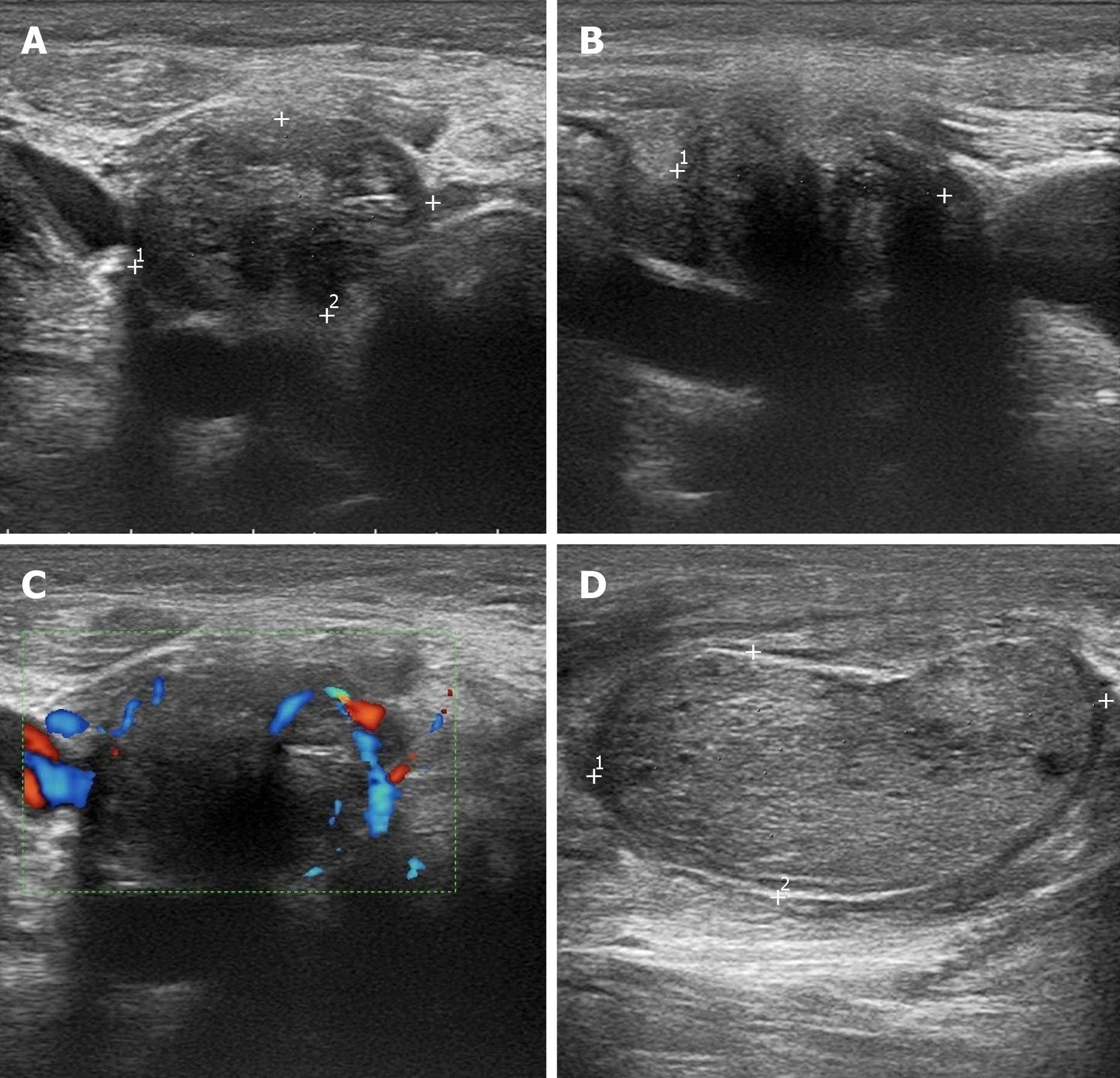
Figure 1 Ultrasound images of calcitonin-negative neuroendocrine tumor with cervical lymph node metastases.
A: Cross-sectional image of the thyroid mass with uneven echo; B: Cross-section image of the thyroid gland with the mass invading the thyroid capsule; C: Color Doppler imaging showing abundant blood supply in the mass; D: Swollen cervical lymph node poorly demarcated between cortex and medulla.
- Citation: Cai HJ, Wang H, Cao N, Huang B, Kong FL, Lu LR, Huang YY, Wang W. Calcitonin-negative neuroendocrine tumor of the thyroid with metastasis to liver-rare presentation of an unusual tumor: A case report and review of literature. World J Clin Cases 2020; 8(1): 179-187
- URL: https://www.wjgnet.com/2307-8960/full/v8/i1/179.htm
- DOI: https://dx.doi.org/10.12998/wjcc.v8.i1.179


