Copyright
©The Author(s) 2020.
World J Clin Cases. Jan 6, 2020; 8(1): 157-167
Published online Jan 6, 2020. doi: 10.12998/wjcc.v8.i1.157
Published online Jan 6, 2020. doi: 10.12998/wjcc.v8.i1.157
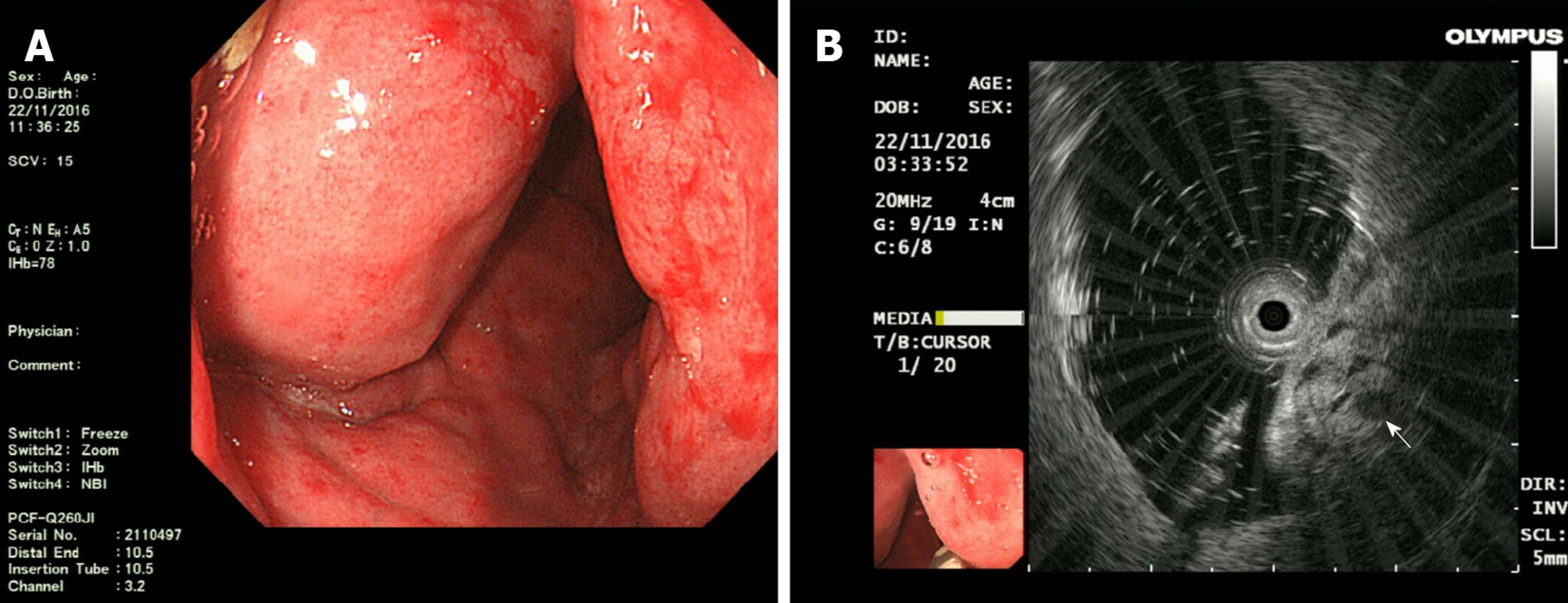
Figure 3 Endoscopic ultrasonography findings: A: Endoscopic ultrasonography (EUS) showed distinct edema and dense vascular varicose dilatations in the submucosal area with erosion and redness in the intestinal mucosa; B: The circumferential wall revealed multiple small “anechoic cystic spaces” on EUS (arrow).
- Citation: Li HB, Lv JF, Lu N, Lv ZS. Mechanical intestinal obstruction due to isolated diffuse venous malformations in the gastrointestinal tract: A case report and review of literature. World J Clin Cases 2020; 8(1): 157-167
- URL: https://www.wjgnet.com/2307-8960/full/v8/i1/157.htm
- DOI: https://dx.doi.org/10.12998/wjcc.v8.i1.157


