Copyright
©The Author(s) 2020.
World J Clin Cases. Jan 6, 2020; 8(1): 110-119
Published online Jan 6, 2020. doi: 10.12998/wjcc.v8.i1.110
Published online Jan 6, 2020. doi: 10.12998/wjcc.v8.i1.110
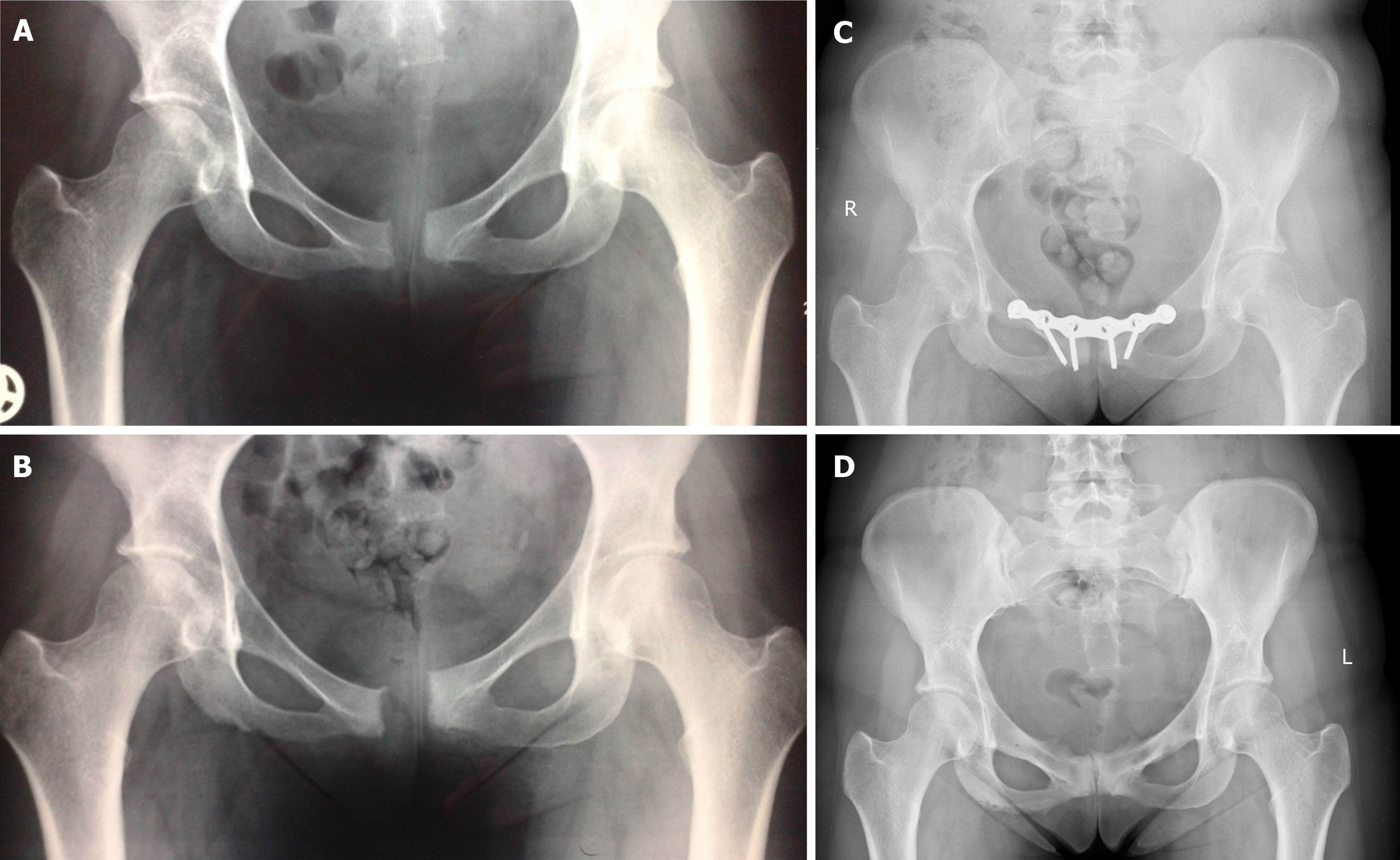
Figure 1 X-ray images of Case 1.
A: 1.5 cm diastasis of the symphysis pubis and signs of secondary pubic osteitis; B: 2.5 cm symphysiolysis, increased signs of pubic osteitis and vertical dislocation of pubic rami; C: 9 mo after initial fixation, no signs of infection, but 4 middle screws are loose. D: 12 mo after hardware removal, signs of pubic osteitis, no signs of instability.
- Citation: Norvilaite K, Kezeviciute M, Ramasauskaite D, Arlauskiene A, Bartkeviciene D, Uvarovas V. Postpartum pubic symphysis diastasis-conservative and surgical treatment methods, incidence of complications: Two case reports and a review of the literature. World J Clin Cases 2020; 8(1): 110-119
- URL: https://www.wjgnet.com/2307-8960/full/v8/i1/110.htm
- DOI: https://dx.doi.org/10.12998/wjcc.v8.i1.110


