Copyright
©The Author(s) 2020.
World J Clin Cases. Jan 6, 2020; 8(1): 11-19
Published online Jan 6, 2020. doi: 10.12998/wjcc.v8.i1.11
Published online Jan 6, 2020. doi: 10.12998/wjcc.v8.i1.11
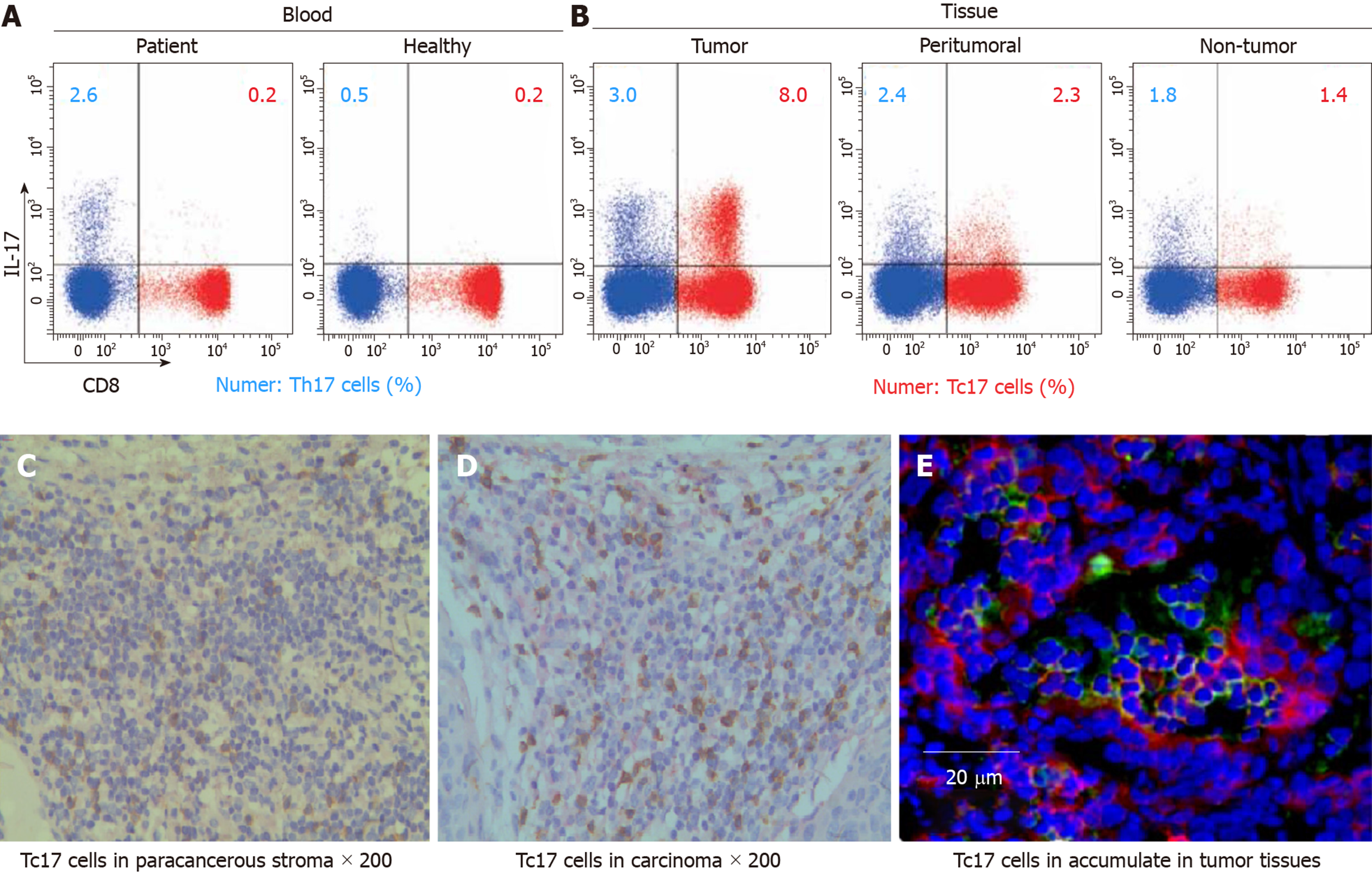
Figure 1 Tc17 cells accumulate in tumor tissues of cervical cancer patients.
A, B: Dot plots of intracellular cytokine staining for Tc17 and Th17 in tumor cells; C: Immunohistochemistry staining for interleukin (IL)-17+ Tc17 cells in paracancerous stroma, the brown signal represents staining of IL-17, and the red signal represents staining of Tc17 (Envision, ×200); D: Immunohistochemistry staining for IL-17+ Tc17 cells in carcinoma nets, the brown signal represents staining of IL-17, and the red signal represents staining of Tc17 (Envision, ×200); E: Immunofluorescence staining for intra-tumoral IL-17+ Tc17 cells, the green signal represents staining of IL-17, the red signal represents staining of Tc17, and the blue signal represents DAPI-stained nuclei (scale bar, 20 μm). IL: Interleukin.
- Citation: Zhang ZS, Gu Y, Liu BG, Tang H, Hua Y, Wang J. Oncogenic role of Tc17 cells in cervical cancer development. World J Clin Cases 2020; 8(1): 11-19
- URL: https://www.wjgnet.com/2307-8960/full/v8/i1/11.htm
- DOI: https://dx.doi.org/10.12998/wjcc.v8.i1.11


