Copyright
©The Author(s) 2019.
World J Clin Cases. May 6, 2019; 7(9): 1053-1059
Published online May 6, 2019. doi: 10.12998/wjcc.v7.i9.1053
Published online May 6, 2019. doi: 10.12998/wjcc.v7.i9.1053
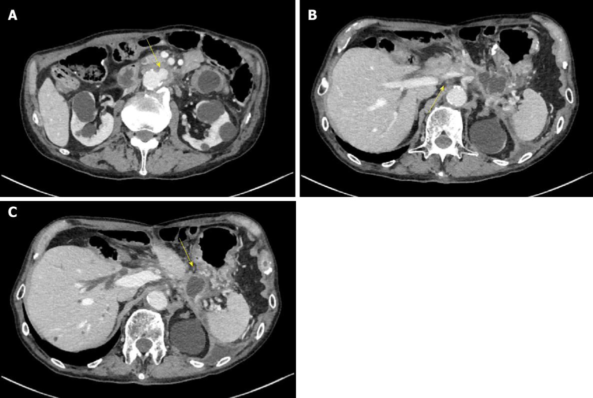
Figure 2 Computed tomography scan taken at 7 years after severe acute necrotizing pancreatitis.
A: Computed tomography (CT) scan showing spread of the pancreatic fluid collection to the aorta with surrounding inflammation and the appearance of ulcer-like blood flow in the aortic aneurysm thrombus (yellow arrow); B: The CT scan also shows splenic vein occlusion (yellow arrow; B) and venous dilation around the residual stomach; C: The shape of the pancreatic fluid collection is narrow around the stomach (yellow arrow), although it shows extensive spread.
- Citation: Yamada R, Umeda Y, Shiono Y, Okuse H, Kuroda N, Tsuboi J, Inoue H, Hamada Y, Tanaka K, Horiki N, Takei Y. Management of the late effects of disconnected pancreatic duct syndrome: A case report. World J Clin Cases 2019; 7(9): 1053-1059
- URL: https://www.wjgnet.com/2307-8960/full/v7/i9/1053.htm
- DOI: https://dx.doi.org/10.12998/wjcc.v7.i9.1053


