Copyright
©The Author(s) 2019.
World J Clin Cases. May 6, 2019; 7(9): 1043-1052
Published online May 6, 2019. doi: 10.12998/wjcc.v7.i9.1043
Published online May 6, 2019. doi: 10.12998/wjcc.v7.i9.1043
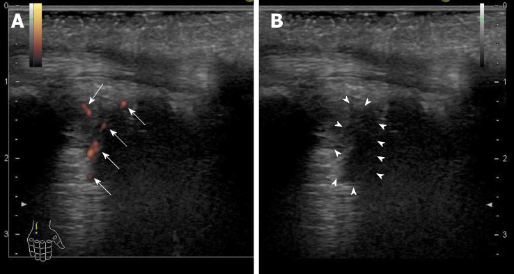
Figure 1 Ultrasonography findings of the right ulnar intercarpal joints in patient 2.
A: Power Doppler imaging. There were clear synovial blood flow signals (arrows) with localization of the synovial thickening; B: B-mode imaging. Synovial thickening was observed as a low echoic lesion (surrounded by arrowheads).
- Citation: Sato K, Yamazaki Y, Kobayashi T, Takakusagi S, Horiguchi N, Kakizaki S, Andou M, Matsuda Y, Uraoka T, Ohnishi H, Okamoto H. Sofosbuvir/Ribavirin therapy for patients experiencing failure of ombitasvir/paritaprevir/ritonavir + ribavirin therapy: Two cases report and review of literature. World J Clin Cases 2019; 7(9): 1043-1052
- URL: https://www.wjgnet.com/2307-8960/full/v7/i9/1043.htm
- DOI: https://dx.doi.org/10.12998/wjcc.v7.i9.1043


